Overview
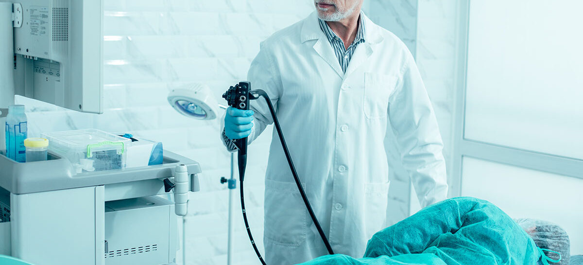
Endoscopy is a popular, minimally-invasive technique used for diagnosis as well as treatment of diseases, in different organ-systems of the body. When it comes to treating disorders of the digestive system (or GI tract), endoscopy is widely used. In this article, we will examine some of therapeutic endoscopy procedures, their purpose and how they are performed.
Introduction
In earlier articles, we have traced the history of endoscopy and covered their usage to diagnose, as well as treat – lung conditions or breathing disorders (Bronchoscopy).
The digestive tract is a 20-25 feet long channel starting at the mouth and terminating at the anus. Various organs and glands in this route are involved in ingesting and digesting food, absorbing nutrients from food, and eliminating undigested waste. Needless to say, any of these organs or parts of the GI tract can develop a condition that requires treatment. While medication is the first course of treatment, this may not work out always. Further, some conditions require more investigation that routine tests cannot uncover.
The availability of two natural openings – the mouth and anus, makes endoscopy a suitable alternative to open surgery. Every year, doctors are innovating newer methods, tools and techniques to not only diagnose (diagnostic endoscopy) but also treat (therapeutic endoscopy) conditions of the GI tract. Here are some of them.
Some therapeutic endoscopies for the GI tract
Endoscopic haemostasis for Ulcer Bleed
Ulcers in the stomach can rupture and bleed. One technique to stop this bleeding is an endoscopic injection of adrenaline into the ulcer and surrounding tissues. In recent times, this technique has been combined with other methods such as hemoclips and heater probe coagulation to reduce chances of the ulcer bleeding again.
Endoscopic Haemostasis for Variceal Bleed
a) Injection sclerotherapy
In people with advanced liver disease, blood flow to the liver gets blocked. This results in enlarged or swollen veins in the lower part of the esophagus. These veins can burst, which is a life-threatening condition. This condition is called esophageal varices. Injection sclerotherapy is a technique that aims to shrink these varices.
b) Variceal banding
Another technique for treating esophageal varices described above. In this technique, bands of synthetic material are placed on the varices. This helps to restrict or cut off blood flow to the veins, thereby preventing bleeding which can be life-threatening.
Argon plasma coagulation
Bleeding is one of the outcomes in conditions like angiodysplasia, GAVE (Gastric Antral Vascular Ectasia) and malignant tumours. Stopping bleeding (called haemostasis) is very important to prevent further complications. Argon plasma coagulation is a technique in which coagulation or clotting is provided to the concerned tissue, thereby preventing bleeding. This is done by passing a jet of argon gas into a catheter that slides inside an endoscope. At the tip of the catheter, an instrument that passes electric current helps ionize the argon gas, which helps to stop bleeding. Since there is no contact between the catheter and the tissue, there is no unwanted tissue damage.
Esophageal dilatation
Achalasia is a condition in which the person cannot swallow properly because of a problem in the esophagus. The lower esophageal sphincter (LES) does not relax properly due to unknown reasons or infection in the myenteric plexus of the esophagus. Esophageal Strictures is another condition in which the esophagus gets narrowed down abnormally. For both these conditions, an endoscopic procedure is done to remove the constriction and make the esophagus wider, which helps to ease swallowing. The technique uses a balloon inserted through a catheter, inflated, and then withdrawn. This method is called endoscopic dilatation.
Stenting
In people with esophageal cancer, the esophagus narrows, so food does not go through smoothly from the mouth to the stomach. This condition is called dysphagia. In such a case, an endoscopy is done in which expandable stents (meshes made of metal) are deployed in the oesophagus, thereby removing any constriction and easing the movement of food. This restores the “Joy of Eating” to patients.
Polypectomy
Gastric/Colonic polyps are unusual clumps or cell-growth on the inner lining of the stomach wall. While most often these growths are not cancerous, sometimes, they may turn into cancer, which is why they must be removed. The procedure of removing them is called polypectomy. Endoscopic polypectomy uses a snare (a ring-like hook) to pull out the polyps, or fulguration that involves heat treatment using hot forceps to destroy the polyps.
Percutaneous endoscopic gastrostomy (PEG)
People who have had a stroke sometimes cannot consume their food in the normal way. They need to be given food through a feeding tube. Inserting this gastronomy tube used to be done surgically, in the past. More recently, an endoscopic procedure called Percutaneous endoscopic gastrostomy (PEG) is used to insert this tube.
Endoscopic foreign-body retrieval
Food boluses while having a meal, and accidentally ingested foreign bodies can get lodged in the lower esophagus, thereby preventing food from moving smoothly. Children most commonly swallow coins or button batteries while adults may swallow dentures accidentally. Endoscopic procedures are used to remove these objects.
Endoscopic resection for lesions
Lesions are abnormal clumps of tissue which may or may not be cancerous. In the stomach and duodenum, such lesions can form on the sub-epithelium or sub-mucosal layers. Endoscopic procedures are done to resection or remove these tissues.
Peroral Endoscopic Myotomy (POEM)
Conditions like achalasia and esophageal spasms can cause esophageal muscles to tighten and make swallowing difficult. POEM is an endoscopic technique to overcome this condition. In this, the endoscopist, cuts the esophageal muscles slightly so that they relax and do not constrict unnecessarily resulting in good symptomatic improvement.
ERCP (Endoscopic Retrograde Cholangiopancreatography)
Procedure done for removing stones stuck in biliary duct or pancreatic duct. It is useful in treating obstructive jaundice / cholangitis.
- A special side viewing scope is used
- The time taken for procedure is 1-2 hours.
- It is done by highly skilled endoscopist.
Kauvery Hospital is globally known for its multidisciplinary services at all its Centers of Excellence, and for its comprehensive, Avant-Grade technology, especially in diagnostics and remedial care in heart diseases, transplantation, vascular and neurosciences medicine. Located in the heart of Trichy (Tennur, Royal Road and Alexandria Road (Cantonment), Chennai, Hosur, Salem, Tirunelveli and Bengaluru, the hospital also renders adult and pediatric trauma care.
Chennai – 044 4000 6000 • Trichy – Cantonment – 0431 4077777 • Trichy – Heartcity – 0431 4003500 • Trichy – Tennur – 0431 4022555 • Hosur – 04344 272727 • Salem – 0427 2677777 • Tirunelveli – 0462 4006000 • Bengaluru – 080 6801 6801


