Summary
Breast Cancer is being increasingly diagnosed in recent years and has caused alarm in women worldwide. Women are being educated about the condition in various forums including our blog-site. Women are being taught how to do a self-examination for symptoms of breast cancer. However, not all women show clear signs and symptoms. This, plus some other factors increases the risk of Breast Cancer in such women. Mammogram is a screening or diagnostic procedure to help detect Breast Cancer early.
Introduction to Breast Cancer
Breast Cancer is the number one cancer in Indian women today, with as many as 25.8 women out of every 100,000 women diagnosed with the condition (age-adjusted figures). Mortality stands at 12.7 for every 100,000 women, with rural women showing more mortality than their urban counterparts (various sources).
Needless to say, early detection makes treatment more effective and minimizes the risk of losing a breast (from a mastectomy procedure). In recent times, there is increased awareness in women across age-groups about the condition, so more and more women are undertaking frequent self-examination of their breasts, today. Self-examination looks for visible signs and symptoms of breast cancer which can trigger timely consultation with an oncologist (cancer specialist) who specializes in Breast Cancer. These signs include swelling and pain in the breasts and nipples, inversion of the nipples, discharge from the nipples, dimpling of breast skin, and red or flaky breast skin.
If and when the woman is experiencing any of these, she can rush to an obstetrician, gynaecologist or oncologist who will do a clinical exam. In this test, the doctor feels the breasts for lumps or growth.
However, there are many women who do not show visible signs listed above or have not felt any lump in their breasts. This puts them at risk, due to a false hope that things are fine. This is dangerous especially if the person has high risk factors for Breast Cancer. In the absence of timely screening, the disease may be advancing silently. A Mammogram is the best solution in such a scenario.
What is a Mammogram
Mammogram, also called Mammography is a clinical procedure in which the woman’s breasts are examined under medium-intensity x-rays. The intensity is lesser than what is used for various diagnostic tests including bone density test. Mammograms are of 2 types:
- Analog mammograms, where the images from the X-ray are captured on a traditional X-ray film. Was the norm till a few years ago.
- Digital Mammogram: Here, X-rays are taken and relayed onto a computer screen, where a software can be used to enlarge or magnify the image in more detail. This again is of 2 types:
- 2D Digital Mammograms: Here a couple of X-ray shots are taken from top, and the sides. All the images are relayed on to the computer screen and can be enlarged for more detail.
- 3D Digital Mammograms: In this, a series of X-ray shots are taken with the machine moving from one side of the breast to the other in an arching motion. The images are relayed to a computer screen. A sophisticated software running on the computer collates these images and creates a 3D picture of the breast. The picture can also be enlarged for more detail.
Who requires this?
There are various risk factors for breast cancer and all such women can benefit from frequent screening using a Mammogram.
- Personal history of breast cancer: Past instances of breast cancer which were successfully treated then.
- Family history of ovarian cancer or breast cancer: Having a mother, grandmother, aunt, sister or daughter who suffered or is suffering from either of these conditions increases the risk.
- Inherited genetic mutations, such as BRCA1 and BRCA2: This is one of the most common risk factors for Breast Cancer.
- Benign or non-cancerous breast conditions such as lobular neoplasia or atypical ductal hyperplasia.
- Dense breasts: Some women have dense breasts which means, there is more fibrous and glandular tissue than fatty tissue. Such women are at higher risk of breast cancer, plus the fact that detecting the condition in them is more challenging.
- Inherited disorders: Where the woman has a parent, sibling or child suffering from Li-Fraumeni syndrome, Bannayan-Riley-Ruvalcaba syndrome or Cowden syndrome
- Radiation exposure to the chest region: Radiation therapy for one or more cancers in the chest region, in the past, increases risk.
When and how often should this be done?
There are 2 purposes for which Mammograms are done:
- Screening Mammograms: This is done on a regular basis, to catch or detect early signs of breast cancer if present.
- Diagnostic Mammogram: If self-examination and clinical examination suspects that something is wrong, a Diagnostic Mammogram is done immediately to confirm or rule out breast cancer.
For the purpose of this article, we will stay focused on Screening Mammograms only.
Women who are 40 years and above are at higher risk. So, age, and risk factors mentioned above will determine the level of risk and hence the frequency of screening Mammogram.
- Low risk: Women who are less than 40 years and do not have any risk factor listed above. Such women can wait till they turn 40 to get their first screening Mammogram done.
- Medium risk: Women who are 40 years and above, and do not have any risk factor listed above. Such women can get a screening every 2 years after the age of 40, and annual screening from 50 years onwards.
- High risk: Women of any age, with one or more risk factors mentioned above. An annual screening is a must.
If you still have doubts, consult an obstetrician, gynaecologist or oncologist at a reputed hospital. He or she will decide the right time to start, and the right frequency, for a screening Mammogram.
How is the procedure done?
Before
- If you are pregnant, or suspect you are pregnant, or a breastfeeding mother, tell the consulting doctor. He/she may postpone the procedure or suggest alternatives such as a breast ultrasound.
- If you are currently going through your menstrual cycle or expecting it shortly, wait for another 2 weeks to schedule a mammogram. This is because the breasts are tender during this interval, so you may experience some discomfort from the procedure.
- If you have been vaccinated recently for one or more diseases, or have a breast implant, tell the consulting doctor. He/she will decide the right time to schedule the Mammogram.
On the day of your mammogram
- Follow all normal routines such as meals, medication, bathing, etc.
- Do not use body powders, deodorants, perfumes or any topical lotion. These appear as white patches on the X-ray images and affect the diagnosis.
- Since you have to be undressed above the waist, wear a two-piece dress (top and bottom). A one-piece dress will require you to undress completely.
During
The procedure lasts around 20 minutes and follows this process:
- You must remove all jewellery and clothes above the waist.
- A healthcare provider will give you an open-front hospital gown, or a drape, to wear.
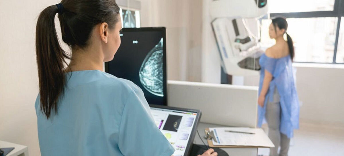
- You must stand in front of the mammography machine and remove one breast at a time from your gown, when you are told to. A lab-technician will position the breast correctly on a breast support plate below the breast.
- The lab-technician will lower a plastic paddle from the top, to compress the breast against the support plate below. This will stretch the breast evenly and horizontally on the support plate, to capture the best possible images. While a slight discomfort is natural, please tell the technician in case you are feeling severe discomfort or even pain. He/she will adjust the pressure accordingly.
- The process is repeated for the other breast.
- Once the technologist is satisfied with the number and quality of images taken, the process is stopped. You will step back from the machine, remove the gown, wear your clothes, jewellery and accessories.
- If test-results are expected immediately, you can collect them and meet the doctor. Else, you will be asked to leave and come later to collect them.
Results, and next steps
There is a Breast Imaging Reporting and Data System (BI-RADS) developed by medical agencies in the west and followed globally today. The score assigned to BI-RADS determines the current situation.
- BI-RADS 0 or ‘Incomplete’: The results are not conclusive, so the doctor may now suggest a Breast MRI or Breast ultrasound to be done for more clarity.
- BI-RADS 1 or ‘Negative’: There is positively no growth or abnormality of any sort. This is what turns out in 90% of the screenings.
- BI-RADS 2 or ‘Benign finding’: There is a non-cancerous growth in the breast such as calcifications, fibroadenomas, lymph nodes or cysts. While this may not be worrisome, the doctor will start a course of treatment for the same.
- BI-RADS 3 or ‘probably benign finding’: There is a 98% probability that this is a benign finding, but that will be confirmed only when a repeat mammogram is done 6 months later.
- BI-RADS 4 or ‘suspicious abnormality’: There is a 2% to 95% probability of the growth being a malignant cancer.
- BI-RADS 5 or ‘highly suggestive of malignancy’: There is more than 95% probability of the growth being a malignant cancer.
- BI-RADS 6 or ‘Known biopsy-proven malignancy’: A previous biopsy done had shown malignant cancer and this score confirms the condition still exists.
With BI-RADS score 4 or 5, the doctor may call for a biopsy to understand the nature of the growth. In all these cases, the doctor will advise the patient on the next steps, if any, that will be taken.
Kauvery Hospital is globally known for its multidisciplinary services at all its Centers of Excellence, and for its comprehensive, Avant-Grade technology, especially in diagnostics and remedial care in heart diseases, transplantation, vascular and neurosciences medicine. Located in the heart of Trichy (Tennur, Royal Road and Alexandria Road (Cantonment), Chennai (Alwarpet & Vadapalani), Hosur, Salem, Tirunelveli and Bengaluru, the hospital also renders adult and pediatric trauma care.
Chennai Alwarpet – 044 4000 6000 • Chennai Vadapalani – 044 4000 6000 • Trichy – Cantonment – 0431 4077777 • Trichy – Heartcity – 0431 4003500 • Trichy – Tennur – 0431 4022555 • Hosur – 04344 272727 • Salem – 0427 2677777 • Tirunelveli – 0462 4006000 • Bengaluru – 080 6801 6801


