3D Imaging of Upper Respiratory Tract
by admin-blog-kh | May 3, 2012 10:22 am
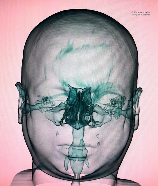
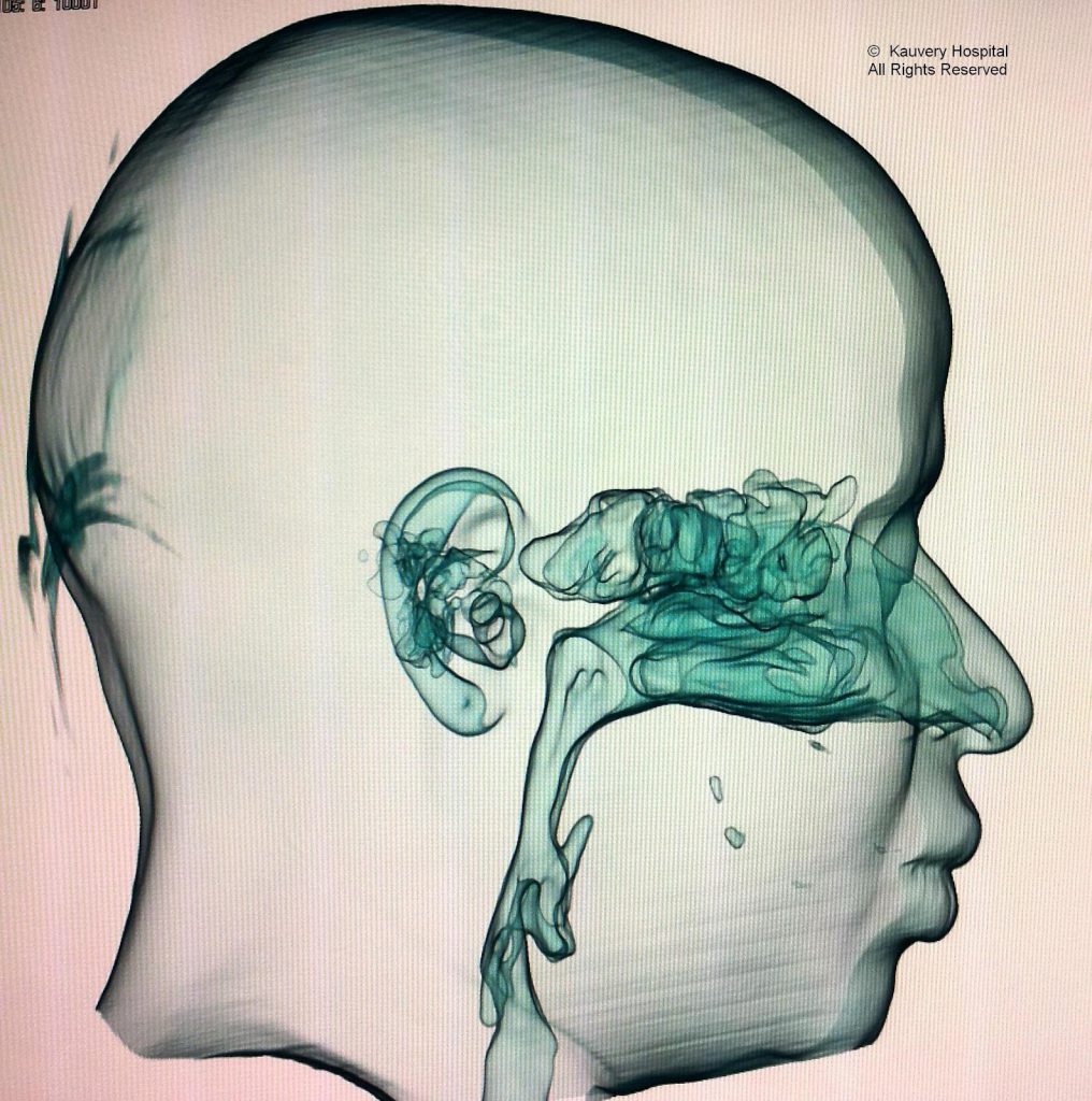
MDCT with volume acquisition enables the 3D reconstruction of complex anatomical regions such as the upper respiratory tract. With this, we can visualise the paranasal sinuses, the nasopharynx, velopharynx, oropharynx and larynx to great detail. For example, in patients with nasopharyngeal lesions, the amount of airway narrowing can be easily visualised and interpreted by the physician[1]. A cine loop of the 3D image can also be made and can be viewed at any workstation. However, it is to be remembered that this is not a replacement for conventional 2D image interpretation, since only air containing regions are visualised here and extramucosal lesions cannot be seen. In the image samples shown below, we can also see the air within middle ear and mastoids.
Kauvery Hospital is globally known for its multidisciplinary services at all its Centers of Excellence, and for its comprehensive, Avant-Grade technology, especially in diagnostics and remedial care in heart diseases, transplantation, vascular and neurosciences medicine. Located in the heart of Trichy (Tennur, Royal Road and Alexandria Road (Cantonment), Chennai, Hosur, Salem, Tirunelveli and Bengaluru, the hospital also renders adult and pediatric trauma care.
Chennai – 044 4000 6000 • Trichy – Cantonment – 0431 4077777 • Trichy – Heartcity – 0431 4003500 • Trichy – Tennur – 0431 4022555 • Hosur – 04344 272727 • Salem – 0427 2677777 • Tirunelveli – 0462 4006000 • Bengaluru – 080 6801 6801
- physician: https://www.kauveryhospital.com/doctors/chennai/family-physician
Source URL: https://www.kauveryhospital.com/blog/radiology/3d-imaging-of-upper-respiratory-tract/