Interventional Radiology Management of Hemoptysis (Coughing up blood)
by admin-blog-kh | April 25, 2012 10:17 am
Massive hemoptysis
Massive hemoptysis (coughing up blood) is a life threatening condition with a mortality rate of 50 to 85 percent with conservative management. embolization. Asphyxiation and less commonly exsanguination are the usual causes of death. Aggressive management is required to manage these emergencies.
Surgical resection of the causative lesion is the initial treatment of choice for those patients with isolated abnormalities and adequate pulmonary reserve.
However, patients with chronic lung disease and limited pulmonary reserve are often considered unacceptable surgical risks, and this group may benefit from bronchial artery embolization.
In addition, there is some evidence that the surgical mortality rate may be lowered by preoperative bronchial artery embolization in those patients who are actively bleeding. It is well established that safe and rapid control of massive hemoptysis can often be obtained by therapeutic transcatheter embolization of the bronchial arteries.
More recently, moderate hemoptysis (greater than or equal to three episodes of 100 ml of blood per day within 1 week) and even mild hemoptysis (chronic or slowly increasing hemoptysis) are considered indications for transcatheter therapy.
Bronchial artery
Bronchial artery is the main source of bleed in patients with massive hemoptysis. However, many non bronchial systemic arteries such as the intercostal arteries, internal mammary artery and inferior phrenic artery may also be the cause for bleed and it is essential to identify these vessels and embolise them for proper management. MDCT with contrast can help in the identification of these vessels and acts as a roadmap for subsequent management.
The embolisation procedure is done by an interventional radiologist[2] and takes approximately one hour time. It is performed in the cathlab under local anaesthesia. The main complication of this procedure is inadvertent embolisation of spinal artery which can result in paraplegia, but this is very unusual.

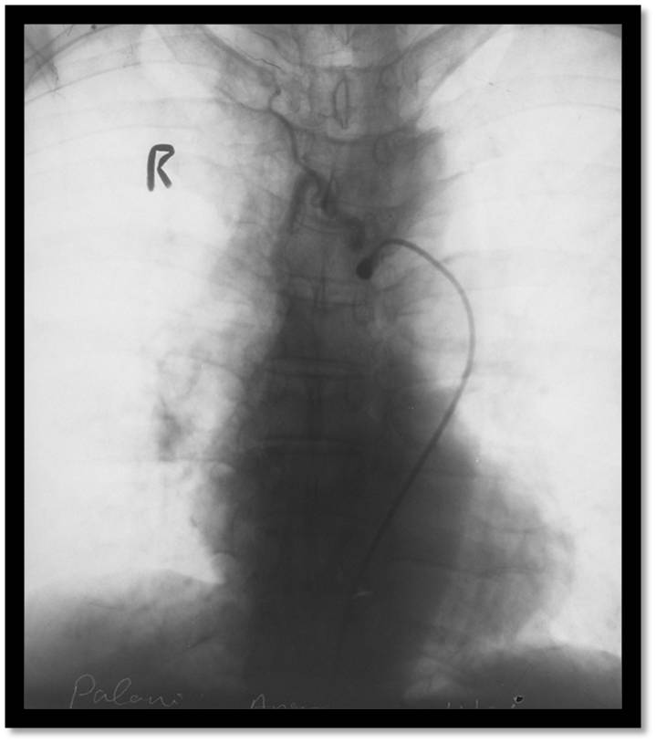
Hypertrophied Bronchial Artery Before Embolisation
Post Embolisation Image Showing Stasis of Contrast
Dr. Iyappan Ponnuswamy[3] MD, FRCR
Consultant Interventional Radiologist, Kauvery Hospital, Chennai
Ph: 044-30606345 | Mob: 09551942599 | Email: [email protected][4]
Kauvery Hospital is globally known for its multidisciplinary services at all its Centers of Excellence, and for its comprehensive, Avant-Grade technology, especially in diagnostics and remedial care in heart diseases, transplantation, vascular and neurosciences medicine. Located in the heart of Trichy (Tennur, Royal Road and Alexandria Road (Cantonment), Chennai, Hosur, Salem, Tirunelveli and Bengaluru, the hospital also renders adult and pediatric trauma care.
Chennai – 044 4000 6000 • Trichy – Cantonment – 0431 4077777 • Trichy – Heartcity – 0431 4003500 • Trichy – Tennur – 0431 4022555 • Hosur – 04344 272727 • Salem – 0427 2677777 • Tirunelveli – 0462 4006000 • Bengaluru – 080 6801 6801
- 3D Imaging of Upper Respiratory Tract: https://kauveryhospital.com/blog/radiology/3d-imaging-of-upper-respiratory-tract/
- interventional radiologist: https://www.kauveryhospital.com/doctors/chennai/radiology
- Dr. Iyappan Ponnuswamy: https://www.kauveryhospital.com/doctors/chennai/radiology/dr-iyappan-ponnuswamy
- [email protected]: mailto:[email protected]
Source URL: https://www.kauveryhospital.com/blog/radiology/interventional-radiology-management-of-hemoptysis-coughing-up-blood/