Introduction
A Felon is a subcutaneous abscess of the distal pulp of a finger or thumb. Superficial infections of the most distal part of the pulp skin are known as apical infections. Apical infections are different from felon in that the palmar pad is not involved. The term Felon should be reserved for those infections involving multiple septal compartments and causing compartment syndrome of the distal phalangeal pulp. The most common organism grown from felons is S. aureus. Gram negative infections have also been reported. Though this condition is uncommon, it is typically seen in immunocompromised.
Case Reports
Case Discussion
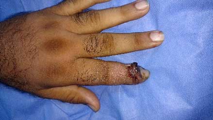
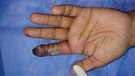
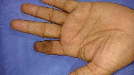
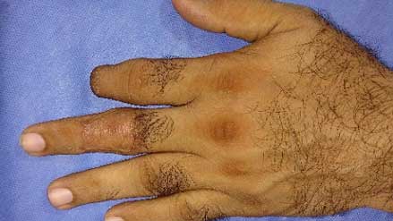
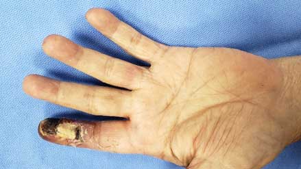
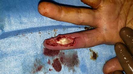
Felons account for 15% to 20% of all hand infections. A felon is characterised by severe throbbing pain, tension and swelling of the entire distal phalangeal pulp. With the progression of swelling and tension there is compromised venous return leading to microvascular injury and tissue necrosis and abscess formation. History of penetrating injury may be there at times. The expanding abscess breaks down the septa and can extend to the phalanx or the skin. If decompression does not occur it is possible that the vessels are obliterated and a slough of the pulp will result. Other complications of an untreated felon include osteomyelitis of distal phalanx, pyogenic arthritis of DIP joint and flexor tenosynovitis and proximal extension.
Treatment
Treatment of felon should be directed toward preserving the function of distal phalanx which includes fine tactile sensibility and stable durable pinch.
In the early cellulitic stage, it is treated with hand elevation and appropriate antibiotics. Immobilization may make the patient more comfortable. Surgical drainage is done by making a unilateral longitudinal incision when there is fluctuance in the pulp. Incisions are made in such a way as to avoid injury to vessels and nerves. Also, the flexor tendon sheath should be preserved. Although it is preferred to avoid placing the incision on the pinching surface, the incision should always be placed on the side of maximal tenderness. When possible, it is made on the ulnar side of the second to fourth digits and on the radial side of the thumb and little finger. The incision is started dorsal to and 0.5cm distal to DIP joint flexion crease. The cavity is enlarged until the adequate evacuation is achieved.
Role of a Plastic Surgeon in Hand Infections
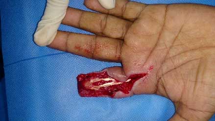
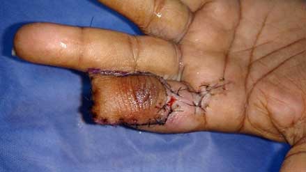
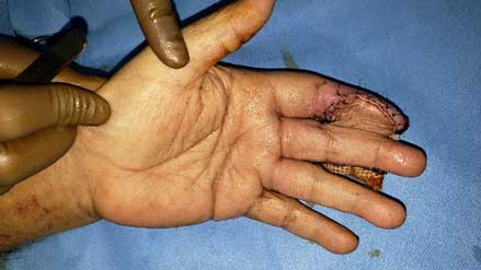
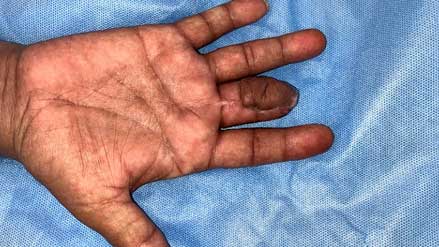
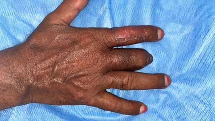
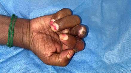
Timely referral of hand infections to a plastic surgeon before compartment syndrome sets in is crucial. Even in the stage of spreading infection, early intervention in the form of debridement arrests further proximal spread. By timely intervention in the form of surgery, we can prevent pyogenic flexor tenosynovitis and septic arthritis of finger joints. In the above cases flap cover was done to preserve the exposed flexor tendon, preserve maximum length of the fingers and to get a functional finger.


