
Endobronchial ultrasound (EBUS) is a technique that uses ultrasound along with bronchoscopy to visualize the airway wall and structures adjacent to it.
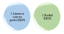
There are 2 types of EBUS:
Indications
- Diagnosing and staging lung cancer
- Sampling hilar and mediastinal lymph nodes of uncertain etiology
- Sampling mediastinal masses
- Draining mediastinal cysts
- Sampling peripheral parenchymal lung nodules(Radial EBUS)
Contraindications
- Severe hypoxia
- Lack of informed consent
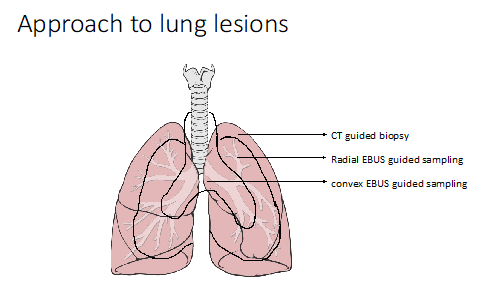
Linear or Convex Probe EBUS
EBUS -TBNA ( Endobronchial ultrasound guided Transbronchial needle aspiration) procedure is done for sampling lesions around larger airways. It needs specialised scope and sampling EBUS TBNA needle or biopsy needle. Sampling is done under real time in this procedure.
 |
 |
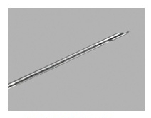 |
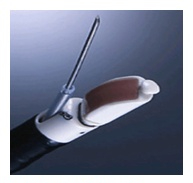
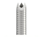
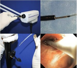
| EBUS scope with needle | Acquire EBUS-FNB needle | COOK ProCore needle |
Case Example:
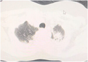
A 32-year-old female patient, who had Covid in May 2021 with no other comorbidities presented with fever, exertional dyspnea, arthralgias and redness of eyes since few months. On examination she had no peripheral lymphadenopathy or organomegaly. Her chest x ray was normal. Her contrast CT chest showed enlarged mediastinal lymph nodes. Her Mantoux was negative, eye examination revealed episcleritis, her calcium and ACE levels were normal. She was taken up for convex EBUS-TBNA
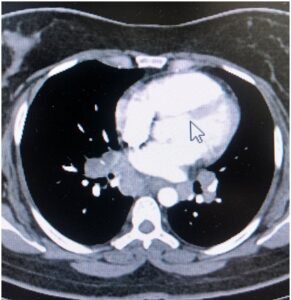
CT chest showing mediastinal lymphadenopathy
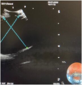
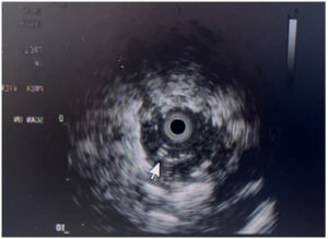
Linear EBUS image showing mediastinal lymph node
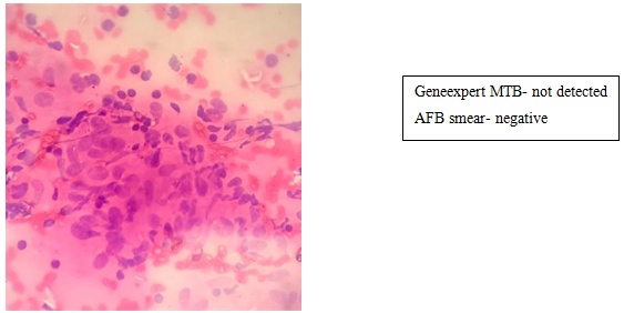
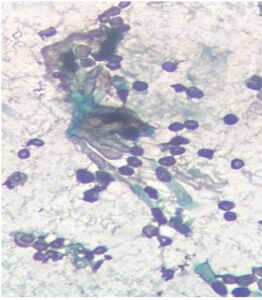
EBUS TBNA sample histopathology slide showing non-caseating granuloma
Final Diagnosis
Sarcoidosis
Table: Comparing diagnostic yield between conventional TBNA and EBUS-TBNA in sarcoidosis (1)
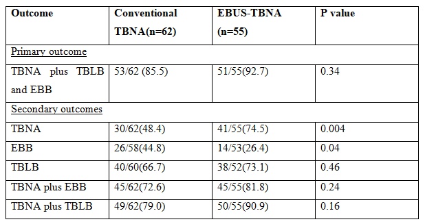
TBNA-Transbronchial needle aspiration
TBLB-Transbronchial lung biopsy
EBB-Endobronchial biopsy
Endobronchial ultrasound guided‑transbronchial needle aspiration vs. conventional transbronchial needle aspiration in the diagnosis of mediastinal masses: A meta‑analysis(2)
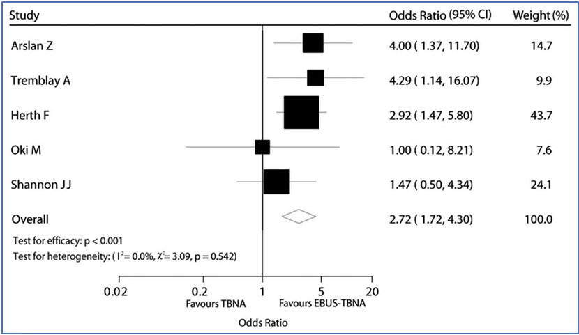
Thus, it becomes clear from the above examples that EBUS-TBNA has more diagnostic yield than conventional TBNA for mediastinal lesions.
It thus can be used in diagnosis of mediastinal lesions like tuberculosis, sarcoidosis and malignancy including staging of cancers
Radial EBUS
Images showing radial EBUS probe generator and radial probe inside flexible bronchoscope
In radial EBUS, a separate radial probe is inserted into the flexible bronchoscope which is then guided into the desired segment/subsegment. The radial probe is then activated giving a 360-degree USG image of the surrounding structures. The target lesion is visualized and sampled. Here, sampling is not done under real time.
Case example:
A 47-year-old female patient who was diagnosed to have crescentic glomerulonephritis in 2020 and on steroids and mycophenolate mofetil for the same presented with cough and weight loss since 2 months. Her CT image showed left upper lobe consolidation with areas of fibro atelectasis and mosaic attenuation bilaterally.
 |
 |
|
CT chest showing left upper lobe |
Radial EBUS image showing the same |
Histopathology slide from Radial EBUS guided BAL/TBLB showing aspergillus fungal elements in fungal stain
The patient was taken up under sedation for Radial EBUS guided TBLB plus BAL which showed aspergillus fungal elements in smear, BAL Galactomannan index of 0.76 and aspergillus flavus growing in culture. The patient was started on voriconazole, and she improved symptomatically over the next 3 months.
Final diagnosis:
Invasive pulmonary aspergillosis
Radial EBUS yield can be further improved by combining it with fluoroscopy/C-arm guidance and VBN(Virtual Bronchoscopic Navigation)
Advantages of EBUS guided sampling over mediastinoscopy
Can be performed under sedation as a daycare procedure, has less cost implications and fewer complications
Summary
Convex EBUS and Radial EBUS have become a game changer in the management of lung lesions. When used in appropriate circumstances they have a lot of advantages like increased diagnostic yield, less morbidity, fewer complications, and lesser cost as compared to conventional techniques.
- Gupta D, Dadhwal DS, Agarwal R, Gupta N, Bal A, Aggarwal AN. Endobronchial ultrasound-guided transbronchial needle aspiration vs conventional transbronchial needle aspiration in the diagnosis of sarcoidosis. Chest. 2014;146(3):547-56.
- Zhu T, Zhang X, Xu J, Tian J, Li H, Liu D, et al. Endobronchial ultrasound guided-transbronchial needle aspiration vs. conventional transbronchial needle aspiration in the diagnosis of mediastinal masses: A meta-analysis. Mol Clin Oncol. 2014;2(1):151-5.
 Dr. R. Nithiyanandan
Associate Consultant Pulmonary and Critical Care
Kauvery Hospital Chennai
