Overview
Several different techniques are used for the closure of surgical wounds. The KENT Team at Kauvery hospital believes in adopting newer methods for a more aesthetically appealing scar, with the use of Cyanoacrylate tissue adhesives to provide a better closing than that achieved by conventional surgical wound closure with sutures or staples.
Cyanoacrylate i.e.-Dermabond Topical Skin Adhesive, (a registered trademark of Ethicon, Fig 2.5) is effective in providing a barrier against the penetration of microorganisms, and also in providing scarless surgery effect. This forms a strong bond across the wound edges that are opposed and aids normal healing.
These are also called liquid sutures and have been proven to reduce closure time. they are flexible as they stay adherent even in dynamic incision sites. They are water-resistant, forming a protective coating over the skin and eliminating the need for suture removal.
The cosmetic outcome with the use of Dermabond is comparable to that of traditional methods of repair. This method of skin closure is best suited for small, superficial lacerations. However, it can also be used with confidence on a larger surgical incision where subcutaneous, buried suturing is needed to approximate the incision site, following which the skin adhesive glue may be used. This method is easy to adopt when patients, especially children, readily accept the idea of not having stitches.
Case Report
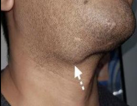
A 50-year male patient came with complaints of swelling over the right side of the neck below the jaw for over 3 years. The size of the swelling had gradually increased over time to attain its present size. It was not associated with pain, restriction in neck movements and presented no pressure symptoms like dysphagia or difficulty in breathing. There was no history of syncope, aspiration or headaches. He only complained of mild pain on palpation and dryness of the mouth on rare occasions.
He had no comorbidities and gave a history of receiving treatment for the condition with oral antibiotics that provided no relief. He was so self-conscious that he began to grow a beard to camouflage the swelling.
 On presentation, he had an obvious swelling of 3.5×4 cms, seen on the right side, over the sub mandibular region. The patient underwent evaluation and a CT scan of the neck was suggestive of a well-defined heterogeneous mass involving the right sub mandibular gland.
The preoperative evaluation was done and surgical fitness was obtained. Surgical excision was planned after a course of antibiotics, which helps in the reduction of gland inflammation. Intra operatively, this aids in the clear identification of planes. Themarginal mandibular nerve identification also is easier in the absence of inflammation. The patient underwent right submandibular gland excision under general anaesthesia. A horizontal skin crease incision was made, blending with the neck crease. The sub fascia approach of the submandibular tumour was used instead of sub platysmal as this helps to reflect the marginal mandibular nerve along with the flap, avoiding nerve injury which may cause neuropraxia or completenerve damage. The hypoglossal nerve, lingual nerve, and the submandibular gland ganglion were identified and preserved, the submandibular duct identified and ligated and the whole right side submandibular gland excised in toto along with its capsule.
Intra operatively, no complications were encountered. Post-operatively, although some patients experience temporary partial marginal mandibular weakness, the complete subsequent recovery of function is usually observed in over one year.

In Fig 2.4, the histopathological section shows salivary gland parenchyma with an encapsulated biphasic cellular neoplasm composed of ductal epithelium cells homomorphically arranged in elongated cords, trabeculae and tubules.
Myoepithelial cells are spindle-shaped, the stroma is chondromyxoid in appearance. Pseudo-cartilaginous areas are seen. Foci show areas of hyalinisation. Peritumoral areas show lymphoid aggregates. Features are suggestive of Pleomorphic adenoma.

Fig 2.6 -2 weeks post-op
Fig 2.7-3 weeks post-op
Discussion
Although salivary gland tumours constitute 1 to 4% of all human neoplasias, they affect:
• The parotid gland in over 70% of the cases
• The submandibular gland in 5 to 10% of the cases
• The sublingual gland in 1% of the cases and
• The minor glands in the remaining 5 to 15% of the cases.
The submandibular gland involvement is seen in only 5% to 10% of salivary gland tumours. Out of these tumours, pleomorphic adenoma is the most common type and this accounts for 90% of all salivary gland tumours. This is followed by the submandibular gland which is in turn followed by the parotid gland. The submandibular gland is the second most common site after the parotid gland. It is frequently the most common benign tumor arising in the submandibular gland.
Pleomorphic adenoma of the submandibular salivary gland is mostly observed in the third and fifth decades of life. Studies have shown the mean presentation of these tumors seen in patients is 44.5 years as observed by Fabio et al. Females are more prone to this than males.
Pleomorphic adenoma characteristically shows a variable amount of myxochondroid stroma produced by myoepithelial cells. The differential diagnosis of basal cell adenoma, adenocarcinoma, mucoepidermoid carcinoma and lymphoma should be considered. However, in a histo pathological study with immuno histochemistry, a patient with mid variabilities in histological grade, mitotic activity and proliferation can be identified This helps when there is a lack of a clear-cut diagnosis of Pleomorphic adenoma which characteristically shows a variable amount of myxochondroid stroma produced by myoepithelial cells.
It presents as a solitary, well-defined, painless, slowly growing benign tumour but can turn malignant. The Ki-67 expression, assessed by immuno histo chemical methods, is used as a key to quantify the mitotic activity, histo logical grade, and proliferation of tumors. Additionally, the mutation of the tumor suppressor gene p53 is the most common genetic cause of human cancers.
A CT scan of the head and neck and magnetic resonance imaging (MRI) of the head and neck are the gold standards of radiological study. Ultrasound guided fineneedle aspiration can be done. Complete excision of the tumor along with the removal of the submandibular gland in toto is the appropriate course of action.
Incomplete removal of the glandular tissue paves the way for a more definitive recurrence of the tumour. About 25% of untreated pleomorphic adenomas undergo malignant transformation. Therefore, early diagnosis and complete excision are recommended. We have taken great interest in giving an aesthetically pleasing scar to our patient- we had done a horizontal skin crease incision which was seen to almost merge and blend into the neck crease. For closure, we used a buried cuticular suture, followed by the use of Dermabond for skin closure.
“An adhesive is a non-metallic substance that adheres or bonds compounds together by adhesion or cohesion without substantially changing the texture of the compounds.” (Definition of adhesives, DIN standard 16920).
The first generation (methyl-cyanoacrylate) is a short-chain cyanoacrylate; this is noted to be histo toxic and can cause foreign-body reactions. The second generation (2-octyl-cyanoacrylate: Dermabond) is a long-chain cyanoacrylate, and is more bio compatible. When it comes in contact with hydroxide ions (liquids like blood or water, air humidity), the 2-octyl cyanoacrylates form long, strong and waterproof chains by an exothermic reaction. This results in a polymer with a stable adhesive bond. The polymerisation time is about 2 min and 20 secs. With excess moisture, the reaction runs too fast for tissue adhesion. Dermabond is approved for clean wounds with easily approximated skin edges. Tissue glue (2-octyl-cyanoacrylate) associated with contact dermatitis is seen inpatients with a known allergy to Formaldehyde, a byproduct of polymerization.
Poor compliance is mostly due to incorrect application and poor wound edge apposition. Repeated crushing of the Dermabond tube can lead to the inner glass vial piercing the outer tube, resulting in a laceration. Hand lacerations can how ever increase the risk of surgical site infection and exposure to blood-borne pathogens.
Hence surgeons should compress the Dermabond tube using forceps and also not repeatedly compress. Sensitization to 2-octyl-cyanoacrylate is considered rare due to the rapid polymerization upon contact with the keratin in the skin. Atemporary inflammatory reaction appears in the glued area. A precondition for functional and aesthetic results is a subcutaneous/subcuticular suture that intercepts the skin tension and helps in the approximation of the skin edges optimally. Bleeding can hamper its application, because the polymerisation would happen too fast for adequate adhesion along with too much heat development.
Tissue glue is contraindicated in infection of the wound, substantial defects, wounds in the range of the eyes and mucous membranes, bites, and very hairy skin.
Conclusion
We describe here our painstaking efforts to give a young man with a sub mandibular tumour an aesthetically pleasing scar, after excision. We have taken this opportunity to discuss the diagnostic and surgical approach to such tumors.
We have also highlighted the benefits of using skin glue in such surgery and also mentioned the possible challenges that may be encountered.
The KENT Team at Kauvery hospital recommends adopting newer methods for amore aesthetically appealing scar, and the use of Cyanoacrylate tissue adhesives to provide a better scar than that achieved by conventional surgical wound closure with suture or staples.


