
Introduction:
Pulmonary hamartomas are the most common benign tumors of the lung, accounting for 75% of all the benign tumors and 8% of all the pulmonary neoplasms. Pulmonary hamartomas present as asymptomatic nodules, found incidentally on routine radiographs and CT scans.
Case Report:
A 56-year-old lady came to OPD for her master health checkup and incidentally discovered a solitary pulmonary nodule on chest XRAY. She was asymptomatic during her examination. High resolution CT scan was performed which confirmed the diagnosis as pulmonary hamartoma.
Imaging Findings:
XRAY:
A plain radiograph was performed in DX (500 mAS) which showed a well-defined round radio opaque lesion in the left hilar region abutting the left heart border
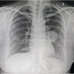
Figure 1: Plain radiograph showing a well-defined round homogenous radio opaque lesion in the left hilar region abutting the left heart border
High Resolution CT:
High resolution CT scan performed in 128 slice GE optima showed a well-defined lobulated hetero dense lesion admixed with areas of fat density in left lower lobe hilar region, measuring 4.8cm x 4.1 cm, suggestive of a pulmonary hamartoma. Few centrinodular opacities were noted distal to the lesion in left lower lobe superior segment possibly due to obstruction of the left lower lobe bronchus by the mass.
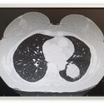
Figure 2: Axial CT showing a well-defined lesion with internal fat density in left lower lobe (-45HU)

Discussion:
Pulmonary hamartomas are the most common benign tumor of lung and is the third most common cause of solitary pulmonary nodule. Patients can be asymptomatic with parenchymal hamartomas, with incidental discovery on chest imaging obtained for any other reason. Hamartomas are composed of fat, epithelial tissue, fibrous tissue and cartilage. Pulmonary hamartomas grow slowly and most of them are smaller than 4cm, although they may reach 10cm in diameter and are usually solitary.
Symptoms:
Pulmonary hamartomas are asymptomatic. Endobronchial hamartomas are associated with obstruction of bronchus with symptoms of fever, cough, expectoration, wheezing and dyspnea.
Radiography:
Pulmonary hamartomas appear as well defined, solitary pulmonary nodule with irregular popcorn, stippled or curvilinear calcification. When calcification or fat is detected in a well circumscribed peripheral lung tumor, diagnosis of hamartoma can be made.
CT Findings:
CT is the diagnostic tool of choice in the study of internal characteristics of pulmonary nodule. The main CT features of pulmonary hamartomas are intranodular fat and popcorn like calcifications.
In HRCT finding of fat and calcification together is specific combination for hamartomas.
Histopathology:
Hamartomas are composed of adipocytes and single chondrocytes appear in lacunae with an abdundant neighboring matrix.
Nuclear Imaging:
Functional imaging with FDG-PET can distinguish between benign and malignant nodules because of increased metabolic activity typically found in cancers.
Treatment:
Patients are usually treated conservatively. Surgical treatment is considered in unresponsive patients and in hamartomas with malignant potential.
Differential Diagnosis:
⦁ Pulmonary chondroma
⦁ Granuloma
⦁ Lung abscess
⦁ Rheumatoid nodule
⦁ Pseudotumor
⦁ Bronchogenic carcinoma
⦁ Pulmonary metastases
⦁ Lymphoma
⦁ Lung cyst
References
⦁ Lundeen KS, Raj MS, Rajasurya V, Ludhwani D. Pulmonary Hamartoma. 2022 Jul 4. In: StatPearls [Internet]. Treasure Island (FL): StatPearls Publishing; 2022 Jan–. PMID: 30969628.
⦁ Leiter Herrán F, Restrepo CS, Alvarez Gómez DI, Suby-Long T, Ocazionez D, Vargas D. Hamartomas from head to toe: an imaging overview. Br J Radiol. 2017 Mar;90(1071):20160607. doi: 10.1259/bjr.20160607. Epub 2016 Dec 12. PMID: 27936889; PMCID: PMC5601532.
⦁ Yamashita K, Matsunobe S, Tsuda T, Nemoto T, Matsumoto K, Miki H, Konishi J. Solitary pulmonary nodule: preliminary study of evaluation with incremental dynamic CT. Radiology. 1995 Feb;194(2):399-405. doi: 10.1148/radiology.194.2.7824717. PMID: 7824717.


