Presentation:
A 32 year old male patient came with C/o swelling in the antero-lateral side of left leg, which appeared on standing, dorsiflexion and disappeared on lying down.
H/o trauma at the swelling site 1.5 years back.
On Examination:
A skin coloured swelling appears on standing and dorsiflexing ankle. The swelling pops up and down while flexing the toes.
Ultrasound:
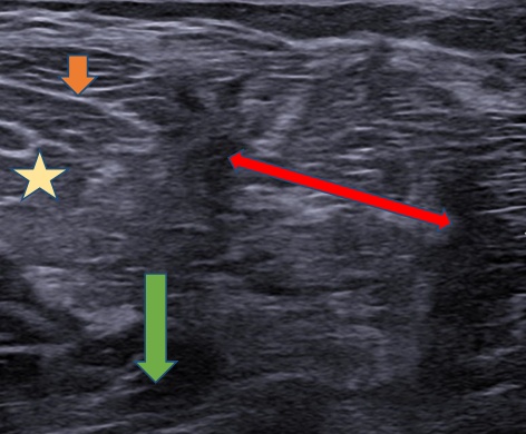
Using 4-16 MHz probe (MyLab 8, Esaote) on the antero-lateral side of the left leg in supine position, a dynamic ultrasound was performed by dorsiflexing his ankle and toes revealed a defect of size in the deep fascia with protrusion extensor digitorum longus in the subcutaneous plane out and in during flexion.


Case Discussion:
A transfascial muscular hernia is a rare type of hernia that occurs when a muscle bulges through a defect in the fascia, a thin connective tissue that surrounds and separates muscles in the body. These hernias can occur spontaneously or after a traumatic injury, such as a sports injury or car accident.
In this case, a patient has presented with a transfascial muscular hernia of the extensor digitorum longus, a muscle located in the lower leg that is responsible for extending the toes, following a trauma that occurred 1.5 years ago. The hernia was detected on a dynamic ultrasound, a specialized imaging technique that uses sound waves to create real-time images of the inside of the body.
Dynamic ultrasound is a useful tool for identifying and evaluating transfascial muscular hernias because it allows for the visualization of the hernia in motion, which can help to distinguish it from other types of hernias or muscle abnormalities. In this case, the ultrasound revealed a small bulge in the fascia of the extensor digitorum longus muscle, consistent with a transfascial muscular hernia.
Treatment for a transfascial muscular hernia typically involves surgical repair to close the defect in the fascia and prevent further protrusion of the muscle. The type of surgery chosen will depend on the size and location of the hernia, as well as the overall health and medical history of the patient. In some cases, minimally invasive laparoscopic surgery may be possible, which involves making small incisions and using specialized instruments to repair the hernia.
It is important for patients with a transfascial muscular hernia to seek medical attention as soon as possible in order to prevent further injury to the muscle and surrounding tissue. With timely and appropriate treatment, most patients can expect to fully recover from a transfascial muscular hernia and return to their normal activities.


