Introduction
Paraneoplastic Rhombencephalitis is a rare and serious neurological condition often associated with underlying malignancy. It is a type of paraneoplastic syndrome, where the immune system generates an autoimmune response against tumor antigens that cross-react with normal neuronal tissues. Paraneoplastic Rhombencephalitis affects the brainstem and cerebellum, regions involved in motor coordination and autonomic functions.
Case:
A 66 years old male presented with progressive imbalance while walking, letharginess hiccups and constipation. CSF analysis was normal.


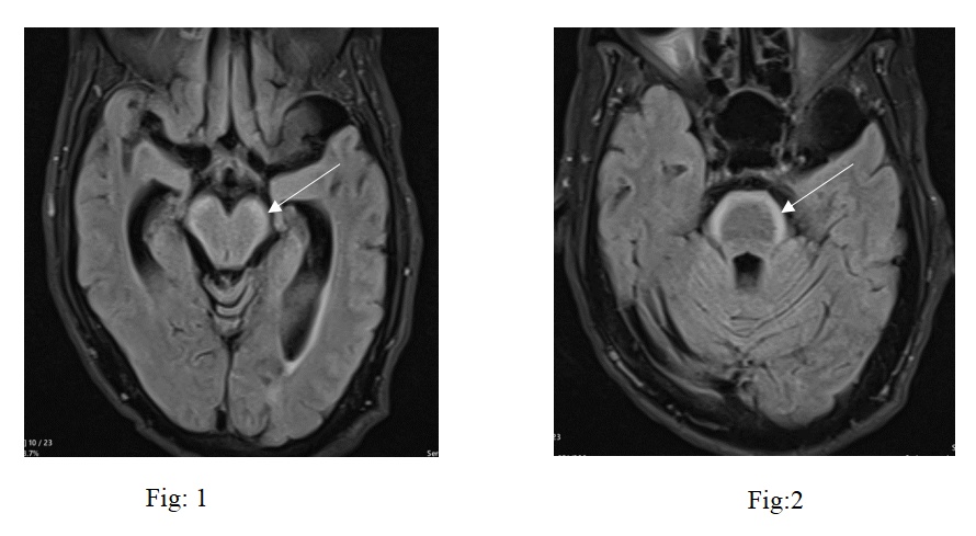
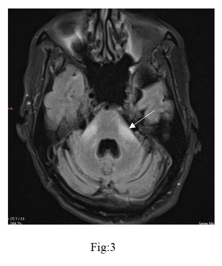
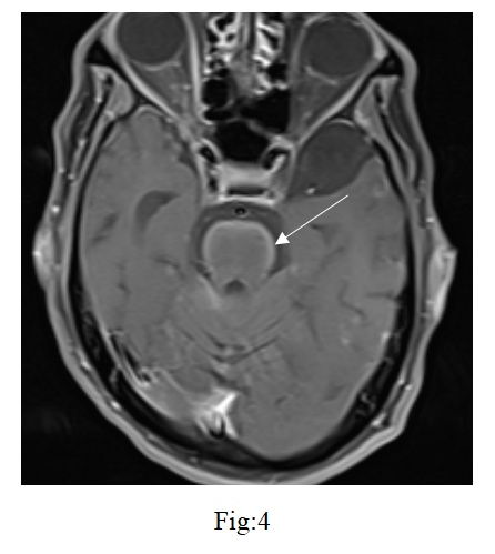
Bilateral symmetric FLAIR hyperintensities in mid brain, pons, medulla and cerebellar peduncles

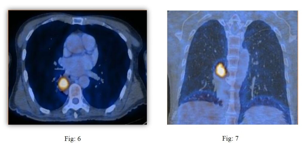
Homogeneous enhancement along the ventral aspect of the midbrain and pons.
FDG avid lesion in superior segment of lower lobe of right lung, Biopsy showed adenocarcinoma
Conclusion:
Paraneoplastic rhombencephalitis (PRE) is a rare but serious neurological manifestation associated with certain malignancies. It results from an immune-mediated response that targets the brainstem and cerebellum and occasionally the spinal cord, leading to a range of neurological deficits such as ataxia, dysarthria, and cranial nerve abnormalities. Advanced imaging, especially MRI and PET scans, is essential for diagnosing paraneoplastic Rhombencephalitis and distinguishing it from other types. It typically has slow progression of symptoms and show predominant involvement of the brainstem (especially the pons and medulla) and cerebellum, with hyperintensities on T2-weighted and Fluid-Attenuated Inversion Recovery (FLAIR) sequences, reflecting inflammatory changes or edema, and while contrast enhancement may be observed in some cases, it is not universally present; in more severe or chronic cases, cerebellar atrophy may develop, contributing to the progression of neurological symptoms, and these radiological findings, when correlated with the clinical presentation of the patient. Particularly a history of malignancy suggestive of a paraneoplastic syndrome, strongly support the diagnosis of paraneoplastic rhombencephalitis, with positive autoantibodies, such as anti-Yo, anti-Hu, or anti-Ri, further aiding the confirmation. This case highlights not only the importance of MRI in picking up brain stem abnormalities not identified in PET scan but also the awareness of primary tumor presenting initially as Rhombencephalitis.
Differentials:
- Infectious Rhombencephalitis: May show focal abscess or nonspecific hyperintensities.
- Autoimmune Brainstem Encephalitis: Have different pattern of involvement.
- CLIPPERS: Shows characteristic punctate and curvilinear enhancement peppering the pons and adjacent structures.
- Neuromyelitis spectrum disorders (NMOSD)
- Myelin oligodendrocyte glycoprotein antibody associated disease (MOGAD).
 Dr. Manish Yadav
DnB Radiology Resident
Kauvery Hospital, Chennai
 Dr. Babu Peter S
Consultant Radiologist
Kauvery Hospital, Chennai
Mentor:
 Dr. Kanagasabai Kamalasekar
Consultant Radiology
Kauvery Hospital, Chennai


