Message from Team IMA Chennai Kauvery Alwarpet Branch

Dear colleagues,
Greetings and Best wishes from IMA Chennai Kauvery Alwarpet branch.
This month’s articles are specially compiled by the Department close to my heart.
The Department Internal Medicine is the pillar of strength in any institution and Medicine is the foundation of any clinician irrespective of their field .
At Kauvery we are a proud Department with five DNB post graduates and the vast clinical exposure they get is evident with articles published. We extend our thanks them for their contributions.
Long Live IMA.
Yours in IMA service,
Dr S Sivaram Kannan
President

Kauvery hospital has been successfully running Medical DNB programmes for the last three years.
This issue brings you articles written by these enthusiastic post-graduates who share their enriching clinical experience.
Long live IMA.
Yours in IMA service,
Dr. Bhuvaneshwari Rajendran
Secretary.

Dear friends
Happy to present this month’s IMA journal to you.
The complete platter in this edition is from the department of Internal Medicine.
We thank all our DNB post graduates for these wonderful presentations.
Kauvery hospitals is proud to nurture such young doctors for their post graduate training who are the health pillars of our future society.
With regards
Dr. R. Balasubramaniyam
Editor

A Case of Myocarditis / Inflammatory Cardiomyopathy
A 56-year-old male, known hypertensive on Tab. Amlodipine 5 mg OD, presented to our emergency department with history of cough with expectoration – mucoid whitish sputum for 2 days, bilateral leg swelling, scrotal swelling and abdominal distension for 2-3 days, decreased urine output for 2-3 days, shortness of breath (NYHA Grade 2 to 3) with orthopnea for 2-3 days. He had history of high grade intermittent fever for 3 days, 1 week back for which he took supportive medications (afebrile for 1 week). He had no history of chest pain, palpitation, syncope, sweating, giddiness, abdominal pain, vomiting, diarrhea, rhinitis, sore throat, myalgia, arthralgia, rashes at presentation or the preceding month. His pulse rate was 90/min, blood pressure- 130/90 mmHg, respiratory rate – 22/ min, SpO2- 99% in room air, temperature- 98F, capillary blood glucose – 122 mg/dl. On examination, he was conscious, oriented to time, place and person, afebrile.

Case of Jejunal Diverticular Bleed
63 years old female known case of diabetes mellitus and systemic hypertension presented with complaints of abdominal pain, loose stools for 5 days. On examination BP – 140/80/PR-86 bpm/SPO2 – 98 percent. Per abdomen examination showed tenderness in umbilical region, no guarding and rigidity. Other system examination were normal. Blood investigation showed elevated WBC counts [14500], CRP [53], RFT and LFT were normal. She was started on intravenous antibiotics and other supportive measures. In the view of persistent abdominal pain CT abdomen with oral contrast was done which showed multiple diverticulosis along the stomach, dueodenum, jejunum, ascending and descending colon. On third day of admission she developed per rectal bleeding which was massive/painless and bright red in colour.

Young Adult with Pancytopenia
24 years old male came with complaints of shortness of breath, loss of weight (5 kg) and appetite, recurrent fever for the past 1 month. Patient had history of giddiness and palpitations for 1 month there was history of generalised fatiguability for 2 months. Family history revealed aplastic anemia/ AML in elder sibling who died at the age of 14 years. There was history of leukemia in cousin who died at the age of 6 years. On examination, patient had tachycardia, pallor and hepatosplenomegaly. Basic blood investigations revealed – Hb:3 g/dl, WBC: 1300/cu.mm (N-40, L-50, M-10, B-0), platelet count: 92000/ cu.mm. Serum LDH level was 405 U/L. RBC indices, renal function tests, live function tests, coagulation profile, vitamin B12 levels, and urine routine were normal. ANA, direct coombs test, CMV IgM, Parvovirus B19 IgM, Hepatitis B and HIV serology were negative. Peripheral smear revealed pancytopenia with more than 20% blasts.

Disseminated Fungal and Becterial Septicemia
Case Report:
A 63 year old male, a known case of type 2 Diabetes Mellitus, Systemic Hypertension and chronic IgG4 pancreatitis for past 3 years on oral steroids presented to ER with complaints of fever on & off associated with productive cough with left eyelid drooping and loss of left eye vision since 10 days. He was diagnosed with IgG4 Pancreatitis on Oct 2020 and started on steroids. On arrival, patient was conscious, oriented, febrile, icteric, tachypneic and tachycardic with bilateral extensive crepitations on auscultation. On examination of left eye, eyelid was drooping and pupil not reactive with extraocular movements full. Patient was admitted in ICU with a provisional diagnosis of fungal pneumonia/ ARDS. CT chest showed focal consolidation with central honeycomb/ bird nest appearance in left UL anterior segment (Fig 1). Patient was started on Inj.

A Case Report of Sequential Post-Covid-19 Vaccine Autoimmune Syndromes (Hyperinflammatory Syndrome, Multifocal Avascular Necrosis, Undifferentiated Arthritis) in Non-Identical Twins – ‘Opening Up of the Pandora’s Box’
The development of any untoward event following vaccination is considered as post vaccine adverse event, though it need not be a causal relationship. Autoimmune syndromes can occur post vaccination with molecular mimicry being the main hypothesis proposed. Similar events have been reported post Covid-19 vaccination and we hereby report a case of non-identical twins (19 year old girl and boy) with serial development of unrelated autoimmune syndromes post booster dose of Covid-19 vaccine (ChAdox1). Initially, four days after the administration of vaccine both developed high grade fever. They were admitted and evaluated for the same. They had pancytopenia with elevated inflammatory markers and all the infectious work up were negative.

Highlights @ Kauvery
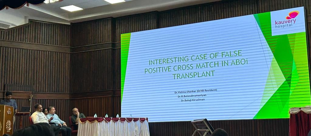
Dr Vishnu our DNB Resident of Nephrology department presented an interesting case of false positive Cross match in an ABO Incompatible Renal Transplant patient from Kauvery hospital in the National Conference organised by Madras Medical Mission Immunology Update. It went well and was appreciated by all the national faculty.

Quiz Of The Month
A 27-year-old female,known case of endometriosis,came with swelling in right inguinal region
Ultrasound
There is a well-defined hypoechoic tubular fluid-filled structure measuring ~ 6.6 x 1.4 cms in the right groin extending laterally along the inguinolabial region. No solid component/ septations noted within the lesion.


