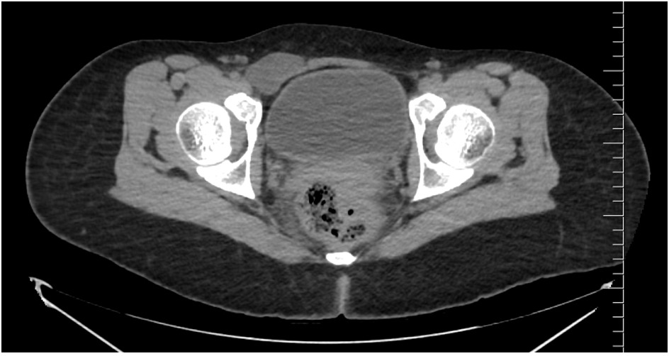
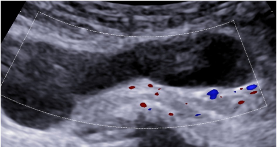
A 27-year-old female, known case of endometriosis, came with swelling in right inguinal region.
Ultrasound
There is a well-defined hypoechoic tubular fluid-filled structure measuring ~ 6.6 x 1.4 cms in the right groin extending laterally along the inguinolabial region. No solid component/ septations noted within the lesion.

On colour Doppler examination – No internal vascularity seen.
On CT Screening

In the right groin is a 6 x 4 cm fluid density structure anterior and medial to the common femoral vein. It has a thin tail extending to the expected location of the superficial inguinal ring.
ANSWER: A case of Hydrocele (Canal Of Nuck)
 Dr Kanagasabai
Consultant Radiologist
