History:
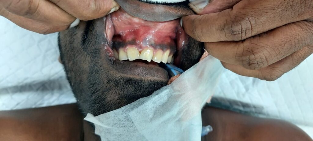
History: Mr A 35 year old patient presented with a swelling in the upper lip as bulging since 1 year. As per the patient he had visited two dentists outside prior to our consultation for dental spacing of front teeth with discolouration. Patient does not remember any injury to the front teeth from childhood or any RTA resulting in any injuries to the teeth.
The dentists after seeing the spacing and discolouration performed radiographs and found the 4 upper maxillary incisors were non-vital and attributed to possible earlier injuries. Root canal treatment was performed on all 4 upper incisors as a first measure. Patient also had very expansible lesion in the anterior maxilla breaching bone and periosteum.
Radiographic and clinical examination confirmed the non-vitality and expansile cyst like lesion in the anterior maxilla. Since they advised surgery patient approached Kauvery maxillofacial unit for further management.
Clinical feature: The swelling in the maxilla was 3×3 cm in size and had no sinus or infection. A CT scan was performed immediately to know the size and extend of the lesion. CT scan confirmed bicortical expansion, thinning of cortical bone and periapical involvement of 3 upper teeth.
Since teeth had only a mild mobility and the lesion appeared benign in nature a decision was taken to remove the cyst and use a filler to fill the bone defect.

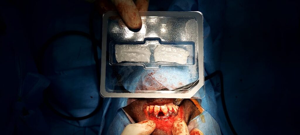
Treatment: under general anaesthesia the patient underwent enucleation of cyst and apicoectomy of roots of 4 teeth. (A process whereby apical root part of the teeth were trimmed and sealed from back of the teeth to prevent the re-entry of infection from cyst). The defect which was nearly 3 cm circumference like a cavity was primarily with filled BMP (bone morphogenic protein) and collagen sponge as a carrier. Additionally allogenic human bone chips were interspersed in to the space for better bone regeneration.

Discussion: Cysts originating from teeth either due to infections or embryonic enamel organ structures. Trauma is another cause for teeth become non-vital and if not rehabilitated like RCT immediately and resulting pulp infection may leads in to cyst formation over a period of time in the bone surrounding. Most these cysts are asymptomatic until expanded cortically not noticeable clinically.
Once a cyst is identified surgeons perform aspiration cytology to know the content of cyst and if cysts are large an incisional biopsy also may be performed. Depend upon the site, cause, age and involvement of facial structures treatment may vary from simple enucleation to complex craniofacial reconstruction. In this case since the cyst may have been originated from periapical infection of teeth and small size in nature a complete primary enucleation was done. Any cyst cavity in the bone larger than 2 cm after operation may produce secondary issues of healing and collapse of the overlying mucosa. Therefore in this case primary regeneration was attempted with BMP and allogenic cortico-cancellous human bone mix. The biopsy of the enucleated cyst confirmed as periapical cyst histologically. Two weeks post-op patient recovered well and advised to do continuous follow-up for up to 6 months.
References:
Nair PN. New perspectives on radicular cysts: Do they heal? Int Endod J 1998;31:155-60.
Shear M, Speight P. Cysts of the Oral and Maxillofacial Regions. 4th ed. Oxford: Wiley-Blackwell; 2007. p. 123-42.
Rajendran R, Sivapathasundaram B. Shafer’s Textbook of Oral Pathology. 6th ed.
St. Louis: W.B. Saunders Elsevier; 2009. p. 273-4.
 Dr. Manikandan Ramanathan
Consultant Maxillofacial Surgeon
Kauvery Hopsital Chennai


