Case history:
A 27-year-old gentleman came to our hospital with chief complaints of swelling and exertional numbness in proximal aspect of left leg. The patient was referred to Radiology for further imaging investigations.
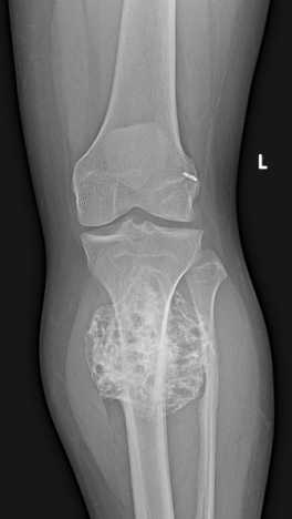
Figure 1 Plain frontal radiograph of left knee
A bony lesion with internal irregular mineralization seen arising from metaphyseal region of left proximal tibia. The articular margins are intact, without any abnormal erosion or sclerosis. No radiological evidence of fracture is seen around left knee. Post ACL repair with endo button in lateral aspect of distal left femur were noted.
MRI of left knee joint:
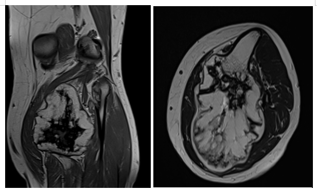
Figure 2A: T1 coronal sequence of left knee joint Figure 2B: T2 axial sequence of left knee joint
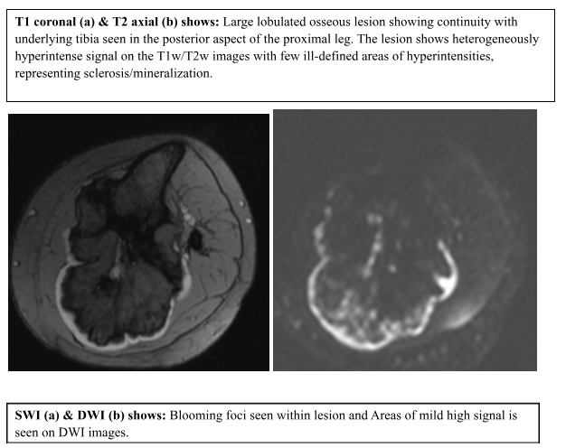
Figure 3A: SWI sequence of left knee joint Figure 3B: DWI sequence of left knee joint
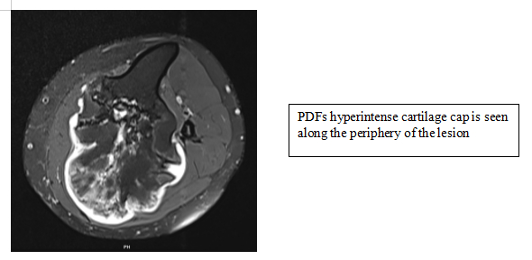
Figure 4: PDF sequence of left knee joint
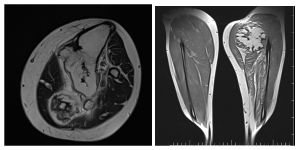
Figure 5A: T2 axial Figure 5B: T1w Cor sequence

DISCUSSION:
Osteochondroma is a benign bony tumor that has a low risk of malignant transformation with an estimated risk of 1% for solitary lesions. Osteochondromas predominantly affect the long bones of the lower extremities, accounting for approximately 50% of cases, followed by knee, femur and tibia
A cartilage cap of over 1.5 cm in thickness after skeletal maturity is suspicious for malignant degeneration. Suspected malignancy in an osteochondroma is often indicated by changes in size or radiographic appearance. The most common form of malignant transformation of osteochondromas is chondrosarcoma.
Complications are a result of the mechanical effects due to their location and size. Vascular complications causing pseudo aneurysm and nerve compression, leading to atrophy of the muscles are seen with larger lesions.
Reference:
- Murphey MD, Choi JJ, Kransdorf MJ, Flemming DJ, Gannon FH. Imaging of osteochondroma: variants and complications with radiologic-pathologic correlation. Radio graphics. 2000 Sep;20(5):1407-34.
- Vasseur MA, Fabre O. Vascular complications of osteochondromas. Journal of vascular surgery. 2000 Mar 1;31(3):532-8.
- Alabdullrahman LW, Mabrouk A, Byerly DW. Osteochondroma. InStatPearls [Internet] 2024 Feb 26. StatPearls Publishing.
 Dr. Kanagasabai Kamalasekar
Consultant Radiology
Kauvery Hospital, Chennai
 Dr. Akshaya Jeyakumar
DMRD Resident
Kauvery Hospital, Chennai
 Dr. Manish Yadav
DnB Radiology Resident
Kauvery Hospital, Chennai


