Case study on Multiple Cranial Nerve Palsy and Necrotizing Pneumonia: The physiotherapy management
Yuvaraj
Physiotherapist, Kauvery Hospital, Salem
Abstract
Multiple cranial nerve palsy refers to a condition where damage occurs to two or more cranial nerves simultaneously leading to dysfunction in various bodily functions like eye movement, facial sensation, swallowing and hearing. It can occur due to number of possible causes,
- Infections
- Inflammatory disease
- Neoplastic disease
- Infiltrative disease of the skull base or meninges
- Idiopathic cause
Necrotizing pneumonia is defined as necrosis of the pulmonary tissue and resulting in the formation of multiple small cavities (<2 cm) containing necrotic debris or fluid. It is a rare, and is a severe complication of bacterial community-acquired pneumonia. Patient who develop necrotizing pneumonia usually have concomitant medical diseases like diabetes mellitus and alcohol abuse. It is characterized by pulmonary inflammation with consolidation, peripheral necrosis and multiple small cavities.
Case Presentation
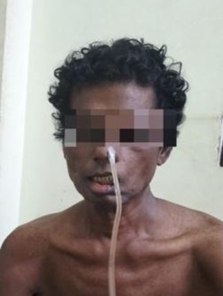
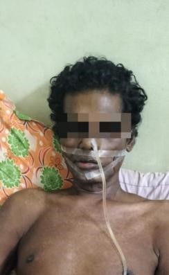
A 52 year aged male presented with history of diplopia from eye deviation followed by swallowing difficulty and chewing, associated with breathing difficulty.
Chief complaints
- Difficulty in breathing
- Difficulty in swallowing and speech
- Difficulty in chewing
Difficulty in eye movements

History
A 50-year-old male, acupuncture therapist from Salem, known to have diabetes and hypertension for the past 7 years, came with complaints of right hemi cranial headache and right-sided earache for the past one month.
He had double vision, on right side more than left side, since 25 days.
He had swallowing difficulty. Bilateral facial weakness for 20-25 days. Unable to close eyes completely. Speech difficulty for 5-7 days.
He was apparently normal before one month.
He had a history of onset of right-sided hemi cranial headache, continuous, that was relieved by medication, after two hours. Double vision on right gaze as well as central gaze, more than left side. Image is more horizontally parted. Diplopia used to resolve after closing either eyes. Onset to peak within 7 days. No redness in eyes or blurring of vision or color desaturation. Since past two weeks, he was having swallowing difficulty, which was static. And it was only for solids as well as liquids, both associated with the regurgitation of food. Bilateral Facial Weakness since 20 to 25 days in the form of unable to close the mouth, unable to close both the eyes, no abnormal sensation or pain, or no history of any ear discharge. Speech difficulty worsening, seen for the past 6 to 7 days, hoarseness of voice and breath is sound, unable to complete the sentence, history of skin rash for the past 10days, no history of any previous history of nausea, vomiting, no history of diurnal variation, no history of joint pain. no history of imbalance while walking, trauma and evil bite vaccination, or recent trauma, no similar type of illness in family.
Past Medical History
The patient has a history of Right-Sided Hemi-Cranial Headache and Right Sided 6th Cranial Palsy which occurred 4 years ago. The condition was treated with Steroids like Ibuprofen, Methylprednisolone, and oral Steroids resulting in improvement within 1 month. Additionally, the patient has a history of Deviation of Mouth to Right Side since childhood. In July 2024, the patient experienced progressive symptoms over 1 month and was admitted to NIMHANS for evaluation and treatment of MNC palsy. The patient received treatment with IVMP and LVPP-2 Cycles made some improvement, and was subsequently discharged with a ryles tube. The patient currently admitted with the same complaint in Kauvery hospital associated with breathing difficulty. History of Diabetes and Hypertension since 6 to 7 years.
Personal history
Non-vegetarian, Not a Smoker, Occasional Alcoholic.
Examination
Patient is conscious, cooperative and well oriented.
Vitals
- Blood pressure – 140/100 mm Hg
- RR – 20/min
- Pulse rate – 100/min
- Spo2 – 98% in room air
Systemic Examination
- CVS- S1, S2 +
- CNS – NFND
- RS – normal
- PA- soft, no organomegaly
Neurological Examination
Hoarse voice and nasal twang
Cranial nerves

- Bilaterally equal and reactive pupil
- Bilaterally isotropic R L
- Bilateral abduction restriction R>L
- Up gaze normal
- Convergence and accommodation present
- Reduced touch sensation on V2 and V3 on right side
- Tongue deviation to Left side
- Uvula deviation to Left side
- Soft palate weak left side
- Bilateral orbicularis oculi weakness +
- Bilateral UMN facial palsy
- Other cranial nerves normal
Motor system examination
- Tone normal in bilateral limbs.
- Muscle Power 5/5 in all limbs
- Reflexes – 2
- Sensory examination Normal
- Cerebellum signs not present
- Skull and spine normal
Investigations reports
1). USG abdomen report on: 11.09.24
Impression: Normal sized bilateral kidneys with raised renal cortical echogenicity. No hydrenephrosis Bilateral mild pleural effusion with partial collapse of right lower lobe
2). Trans-Thoracic Echo report on: 11.09.2024
Impression: Mild concentric LVH/ Mildly dilated LA/ No RWMA Grade I LV diastolic dysfunction, Trivial mitral regurgitation, Mild tricuspid regurgitation, Mild pulmonary artery hypertension, No clot/vegetation/ pericardial effusion
3). MDCT chest plain Report on: 13.09.24
Impression: Diffuse air space ground glass opacities in dependent portions of right lower lobe and entire left lower lithe M Few patchy sub pleural consolidation with central cavitation’s in both lower lobes.
- Necrotizing pneumonia
- Diffuse mosaic attenuation in both lungs
- Lobulated pleural effusion in left upper lobe with fissural extension Focal bronchiectasis with peri-bronchial thickening in right middle lobe
- CORADS-2.
High-resolution ultrasound of right forearm (bedside) report on 14.09.24
Impression: Focal irregular echogenic collection measuring 25 x 15 mm seen in antero lateral aspect of right mid forearm abutting cephalic vein, Visualized cephalic vein in forearm is thrombosed, Ill-defined hypoechoic collection seen in subcutaneous plane of proximal and mid forearm in lateral aspect measuring 10 mm .No internal color flow seen within the collections
Hematoma.
Histopathology report on: 19.09.24
Tissue with focal suppurative inflammation submitted biopsy is negative for fungus
Diagnosis
- Sepsis/Necrotizing pneumonia/Tinea versicolor
- Multiple cranial nerve palsy- Immune mediated (VI,VII,XII Cranial nerve)
Physiotherapy Management
Goals
- To improve facial expression, swallowing, chewing, speech, eye movements
- Reduce breathing difficulty
- Make the patient ask to performing ADL comfortably
Electrical stimulation
The electrical stimulation given 30 contractions each muscle groups
- Muscles of face: Galvanic stimulation
- Nerve trunk: Faradic stimulation
- Muscle of pharynx: Galvanic stimulation

Exercise
- VI cranial nerve palsy
- Cawthrone-cooksey exercise (10 times every 2-3 hr

In bed or sitting
- Eye movements – at first slow, then quick up and down from side to side focusing on finger moving from 3 feet (1 meter) to 1 foot (30 cm) away from face.
- Head movements at first slow, then quick later with eyes closed. Bending forward and backward, turning from side to side, turning head up and down.
In sitting
- Continue the eye movements
- Shoulder shrugging and rotation
- Bending forward and picking up objects from the ground.
In standing
- Sit to stand with eyes open and closed
- Throwing a ball above eye level from hand to hand
- Changing from sitting to standing and turning around in between
Moving about - Circle around centre person who will throw a large ball and to whom it will be returned
- Walk across room with eyes open and then closed
- Walk up and down slop and steps with eye open and closed
VII Cranial nerve palsy Facial exercise 10×3 (3 times a day)

Facial Massage – Stroking, Effleurage, Picking up, Kneading, Rolling

Facial tapping: At night

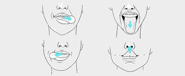
XII cranial nerve palsy (10 reps per hour)
- Tongue pushups
- Tongue curls
- Tongue twisters
- Mewing

Breathing exercise
- Glossopharyngeal breathing (15 times every 2-3 hrs)
- Spirometry (15 times every 2-3 hrs)
- Pursed lip breathing (15 times every 2-3 hrs)
- Chest physio (percussion) 3 times a day
- Huffing and cuffing (15 times every 2-3 hrs)
- Diaphragmatic breathing (15 times every 2-3 hrs)
Advice for recovery
- Exercise regularly
- Apply moist heat to the paralyzed areas to help reduce pain
- Massage affected area
- Tape the eye closed for sleeping
- Use protective glasses or clear eye patches to keep the eye moist and to prevent foreign materials from entering the eye
Outcome Measure
1. House Brachman scale Grade Description
- Pre Treatment analysis on 20/09/2024 showed Grade 5
- Post Treatment analysis on 04/10/2024 showed Grade 2
2. Grading of Force Duction Test
- Pre Treatment analysis on 20/09/2024 showed Grade 3+
- Post Treatment analysis on 04/10/2024 showed Grade 2+
3. Functional grading of dysphagia
- Pre Treatment analysis on 20/9/2024 showed Grade 6
- Post Treatment analysis on 04/10/2024 showed Grade 2
Conclusion
Pre and posttest values, this study concludes that electrical stimulation with exercise found to be effective in improving swallowing, chewing, facial expression, and reducing breathing difficulty in multiple cranial nerve palsy associated with Necrotizing pneumonia.

