Cardiac’s Myxoma
Logeswari B
Cardiac Technician, Heart City, Kauvery Hospital-Trichy.
Background
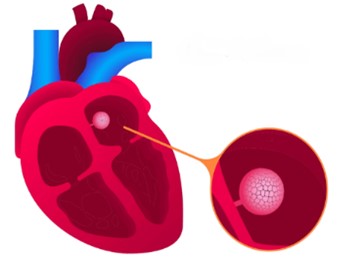
A myxoma is a benign (non-cancerous) tumor of the heart. Myxomas can be as little as one centimeter across or as big as fifteen centimeters. The left atrium, or upper left chamber of the heart, is where the majority, or almost 74%, develop. The right atrium is where the remaining 18% happen. Because these tumors are located in the upper chambers of the heart, they are usually referred to as atrial myxomas. The ventricles, the heart’s lower chambers, are where the remaining 8% grow. Myxomas may sprout from the septum, the wall that divides the heart into two halves. They are frequently joined to this wall by a pedicle, which is a stalk that gives the tumor mobility. Despite not being malignant, myxomas can cause problems for the heart, which makes them dangerous.
Pathophysiology
There are other shapes that myxomas can take, such as polypoid, spherical, or oval. Their surfaces might be lobe-like or smooth, and they resemble jelly. They usually have a white, yellowish, or brown appearance. They can originate from the atrial appendage, the front of the atrial wall, the rear of the atrial wall, or the margin of the fossa ovalis in the left atrium, which is where they are most frequently encountered. The tumor’s stalk length and degree of attachment to the interatrial septum affect how much movement it can make. These characteristics are typically present in sporadic myxomas, which are isolated tumors. On the other hand, familial myxomas that run in families can originate in strange areas, be found in several locations, and form a cluster. Additionally, myxomas rarely impact both heart valves, while they occasionally can affect more than one heart chamber. As a component of the Carney complex, this is possible. Familial myxomas can be detected in more than one location, but sporadic myxomas are usually identified in just one.

Fig.1: Myxoma in the Left Atrium
Causes
Non-malignant tumors happen when cells in the body multiply and split more than they should, without spreading into nearby tissues. Typically, the majority of myxomas happen randomly, indicating that there’s often no specific reason for their development.
Risk factors
Certain risk factors increase chances of developing an atrial myxoma, including:
Being female significantly raises the risk of myxomas, as they are more prevalent in women and may be linked to heart valve issues and the irregular heartbeat known as atrial fibrillation.
Age plays a role too, with the highest risk period being between 20 and 40 years old.
Family history matters: there’s evidence linking some myxomas to Carney’s complex. This is an uncommon genetic disorder that leads to the growth of benign tumors in children and young adults. Since 1985, approximately 750 individuals worldwide have been diagnosed with this condition.
Symptoms
Many individuals affected by myxomas do not show any signs, but a few do. The signs of a myxoma differ depending on its position inside the heart.
- Lethargy (lack of energy).
- Night sweats.
- Breathing difficulty when lying flat or on one side or the other.
- Chest pain or tightness.
- Dizziness and fainting.
- Sensation of feeling your heart beat (palpitations).
- Shortness of breath with activity.
Complication
Complications may include:
- Arrhythmias
- Pulmonary edema
- Peripheral emboli
- Blockage of the heart valves
Diagnosis in echocardiogram
During an echocardiogram, a myxoma shows up as a heterogeneous, movable lump with one of the two main types. Polypoidal myxomas are bigger, featuring a bumpy and smooth exterior and a rough core including lucencies and cystic areas due to hemorrhage and necrosis. In contrast, papillary myxomas are smaller and appear stretched with many tiny projections. The second type is linked to embolic phenomena, while the first type blocks the heart’s blood flow, leading to potential heart failure signs.
Transthoracis echocardiography (TTE)


Fig.2: LA Myxoma in 2D-ECHO

Fig.3: RA Myxoma in 2D -ECHO
Two-dimensional (2-D) echocardiography is frequently sufficient for reaching a diagnosis, even though TTE is not more sensitive. Tumor features including position, size, connectivity, and mobility can all be assessed using this method. Tumors can occur anywhere in the heart, therefore it’s critical to check all four chambers. A left atrial thrombus can be distinguished from an atrial myxoma by its layered appearance and characteristic location towards the rear of the heart. A stalk and mobility are characteristics that differentiate an atrial myxoma from a thrombus.
Doppler echocardiography
Assessing the heart’s chamber pressures and blood flow velocity is a crucial component of Doppler echocardiography when considering cardiac myxoma. This information can be useful in assessing the health and functionality of the heart valves. This method can demonstrate how atrial myxoma affects the dynamics of blood flow. Sometimes it’s combined with contrast-enhanced ultrasonography, which uses contrast agents in the form of gas-filled microbubbles to improve imaging of fluid motion characteristics like blood flow rate.
M-Mode
Using the M-mode echogram technique can be useful for examining irregular echoes in the atrial space. Due to space constraints, it might not be possible for an ultrasound wave to pass through a tumor, especially if it’s small or if the tumor stays inside the atrial cavity.

Fig.4: LA myxoma in M-Mode
Treatment
The surgery is the preferred method of treatment; the tumor growth must be eliminated through surgery. Additionally, certain individuals might also require the replacement of their mitral valve during the procedure. This procedure can be completed simultaneously. Typically, failure of completely removal of the tumor growth originating from a metastasis or implantation of the primary tumor within the heart are common reasons for a recurrence of the tumor and it tends to happen within the first 10 years following surgery, particularly among younger individuals.
Prognosis
Although these tumors are benign and may not spread to other parts of the body, they often cause various health issues. Without treatment, these tumors may rupture (tumor cells are dislodged and carried by the blood) and can end up in vital areas like the brain, eyes, or arms and legs. There might also be issues with the heart as the tumor grows, potentially blocking blood through the valves, leading to conditions such as valve stenosis (narrowing of the valve) or valve regurgitation (blood flowing backward through the valve). This situation could necessitate urgent heart surgery to avoid sudden cardiac arrest.
Case Presentation
Successful Excision of Left Atrial Myxoma
A 34 Years old male patient presented with following complaints
- Dyspnea (shortness of breath)
- Fever
- Night sweats
- Swelling in the legs
Medical History: Diabetic and Normotensive
Diagnosis
After a thorough evaluation, patient was diagnosed with a Left Atrial Myxoma, a rare heart tumor.
Pre-operative ECHO image and report


Treatment
Consultants explained the severity of the condition and recommended surgical excision. The patient provided informed consent, and the surgery was successfully performed on May 31,2024.
Post-operative care and outcome
During his post-operative stay, he received necessary care and monitoring. His condition improved steadily, and he was discharged on June 5,2024, with a stable hemodynamic status.


Conclusion
This case highlights the successful management of a rare cardiac condition, LA myxoma, through timely surgical intervention and appropriate post-operative care.

