Cardiac Tissue Viability Study
T Jasmine Rajareegam Princely1, T Ambika2, G Nagalakshmi3
1Non Critical Ward Incharge, Kauvery Heartcity, Trichy, India
2Senior Staff Nurse, Kauvery Heartcity, Trichy, India
3Junior Staff Nurse, Kauvery Heartcity, Trichy, India
*Correspondence: +91 8508698000; nursing.heartcity@kauveryhospital.com
Abstract
A Cardiac Viability Scan is done to assess how much the heart muscle has been damaged by a heart attack or heart disease. It checks blood supply to the heart and how well the heart muscle works.
Background
- Cardiac MRI: MRI is much more accurate than both nuclear and echo stress, and a cardiac stress MRI provides more information than perfusion, including viability, function and morphology, at a much higher resolution than either nuclear or echo.
- A thallium stress test is an imaging test that indicates how well blood flows into your heart while you ‘re exercising or at rest. It ‘s also called a nuclear stress test.
During the procedure, a small amount of thallium, a radioactive tracer, is administered into a vein on the arm. The tracer is a dye that makes blood flow visible to a special camera called a gamma camera. This camera can reveal any issues the heart muscle may be having.
The test includes exercise and resting portions; the heart is photographed during both.
Single Photon Emission Computed Tomography (SPECT) is a non-invasive procedure that can accurately identify areas of abnormal myocardial perfusion, determine the functional capacity of your heart muscle and separate viable (living) from non-viable (irreversibly damaged) tissue.
Case Presentation
A 50-years aged patient, with a history of diabetes and coronary artery disease (CAD). His angio had earlier shown double vessel disease (17/08/2022), had presented with intermittent chest pain for one week.
Patient initially consulted a hospital and was referred here for further management.
On examination, the patient was conscious and oriented and vitals were stable. ECG and Echo indicated moderate to severe LV disease.
In view of the above findings, the patient was advised admission for further management. He was started with optimal cardiac medications. Medical stabilization was done. The patient was planned for a tissue viability study.
The need for revascularization had to be decided after the viability study through Cardiac MRI or Stress Thallium. It was planned to proceed with PTCA if the LAD territory seemed viable and planned for medical management until then. His cardiac medications were titrated.
At the time of discharge, Echo revealed Severe LV dysfunction with LV auto contrast hence the patient was started on triple anti-platelets. The patient was discharged in a stable state.
Vital signs
Temp – 98.6F, Pulse -88bpm, Resp -22bpm, B.P -130/90 mm of Hg,
Urine Output was adequate.
ECHO
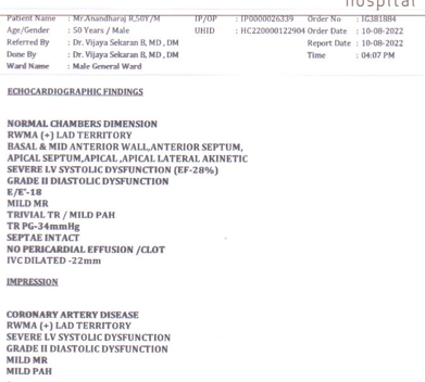
X-Ray Chest: Within normal limits
Nursing Management
- Explain about the procedure to the patient.
- Provide psychological support.
- Maintain interpersonal relationship with patient and family members.
- Diversional therapies like newspaper reading, listening to music was introduced.
- AIDET communication technique was followed for effective communication.
- Bundle chart was assessed updated every day.
- Strict compliance on HICC standard precautions was ensured.
- The nurses assessed the skin integrity of the patient on a daily basis to prevent dehydration or skin tear.
- Necessary investigations were done on a daily basis to monitor his health improvement.
- The assigned nurses communicated with the patient reassuring him about his health status and gave him orientation to time, place and person.
- Nurses coordinated with other health care professionals for care while doing X ray, arranging diet at appropriate time and for other timely investigations.
- Nurses encouraged and engaged him in active and passive exercises like deep breathing exercise.
- They coordinated his physiotherapy as well.
- Advised and observed for not straining on defecation to prevent dyspnoeic and hypertensive crisis.
- Nurses positioned him two hourly, round the clock and provided back care and back massage like effleurage, petrissage, tapping and friction, for prevention of bedsore. Each nurse communicated effectively and developed good rapport with the patient, attender and he cooperated well in his care.
- Nurses developed a home like environment for him to relax and encouraged him to sit on the chair with O2 support, every day.
Conclusion
Patient was in a hemodynamically stable status and was discharged with advice to proceed with Tissue Viability Test. Patient ‘s prognosis was explained in detail.

Ms. Jasmine Rajareegam Princely
Nursing Incharge

Ms. Ambiga
Senior Staff Nurse

Ms. Nagalakshmi
Junior Staff Nurse