Clipping of a Cerebral Aneurysm at Nellai
Jeya Karunya
Staff Nurse, Kauvery Hospital, Tirunelveli.
Abstract
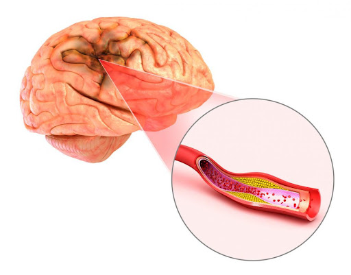
An aneurysm is an enlargement of an artery caused by weakness in the arterial wall. Often there are no symptoms, but a ruptured aneurysm can lead to fatal complications. An aneurysm refers to a weakening of an artery wall that creates a bulge, or distention, of the artery. Aneurysms often occur in the aorta, brain, back of the knee, intestine or spleen. A ruptured aneurysm can result in internal bleeding and stroke. It can sometimes be fatal. Aneurysms often have no symptoms until they rupture. Treatment varies from watchful waiting to emergency surgery. The choice depends on the location, size and condition of the aneurysm. Aneurysms have a variety of causes including high blood pressure and atherosclerosis, trauma, heredity, and abnormal blood flow at the junction where arteries come together. Although the exact cause of an aneurysm is unclear, certain factors contribute to the condition. For example, damaged tissue in the arteries can play a role. The arteries can be harmed by blockages, such as fatty deposits. These deposits can trigger the heart to pump harder than necessary to push blood past the fatty buildup. This stress can damage the arteries because of the increased pressure. Mycotic aneurysms are caused by infections of the artery wall. Tumors and trauma can also cause aneurysms to form.
Background
The patient was a 65-years-aged female. She was diagnosed with cerebral aneurysm (CA) -3.0*1.3 and 3.0 *1.5 mms. After history taking, and examination the consultant, the plan was to offer her Bifrontal craniotomy, clipping of left sided aneurysm and wrapping of the right side aneurysm from left A2.
Examination
- CVS: S1, S2, RS: Bilateral air entry is present, P|A: soft, CNS: NFND
- Temperature: 98.6 F, SaO2: 90%, BP: 160\90, HR: 60beats\mts, RR: 14breath\mts
Investigation
CT Scan: Subarachnoid hemorrhage, diffuse cerebral edema.
CT Angio Scan: Bi-lobed aneurysm seen arising from A3 segment of left anterior cerebral artery. The aneurysm is 3.0*1.3 and 3.0*1.5 mms.
Provisional diagnosis
- Subarachnoid hemorrhage
- Diffuse cerebral edema.
- Bilobed left A3 segment aneurysm.
Surgery
Craniotomy was done under general anesthesia. Aneurysm was clipped. Continuous monitoring was done of the patient. Prescribed antibiotics, analgesics and other medications were given. GCS improved. Healthy surgical wound was seen.
Treatment
- Inj.Cefriaxome-1 gm
- Inj.Levipil-1 gm
- Inj.3%Nacl-6 hourly once
- Inf.Nitroglycerin-2 ml/hr on flow
- Inj.Midazolam
- Inj. Mannaitol-6 hourly once
- Tab.Nimodipine-60mg
- Tab.Cilacor-10 mg
- Syp.cremaffin-10 ml.
Diet advice:
During post-operative periods, we initiated liquid diet. After that, semi solid, solid diet and normal diet were given to the patient and was tolerated.
Nursing management:
Improving Cerebral Tissue Perfusion
- Monitor closely for neurologic deterioration, and maintain a neurologic flow record.
- Check blood pressure, pulse, level of consciousness, pupillary responses, and motor function hourly; monitor respiratory status and report changes immediately.
- Implement aneurysm precautions (immediate and absolute bed rest in a quiet, nonstressful setting; restrict visitors, except for family).
- Elevate the head of the bed 15 to 30 degrees or as ordered.
- Avoid any activity that suddenly increases blood pressure or obstructs venous return (eg, Valsalva maneuver, straining), instructs the patient to exhale during voiding or defecation to decrease strain, eliminate caffeine, administer all personal care, and minimize external stimuli.
- Apply anti-embolism stockings or sequential compression devices. Observe legs for signs and symptoms of deep vein thrombosis tenderness, redness, swelling, warmth, and edema.
Relieving Sensory Deprivation
- Keep sensory stimulation to a minimum.
- Explain restrictions to help reduce the patient ‘s sense of isolation.
- Relieving Anxiety
- Inform patient of plan of care.
- Provide support and appropriate reassurance to patient and family.
Monitoring and Managing Potential Complications
- Assess for and immediately report signs of possible vasospasm, which may occur several days after surgery or on the initiation of treatment (intensified headaches, decreased level of responsiveness, or evidence of aphasia or partial paralysis). Also, administer calcium channel blockers or fluid volume expanders as prescribed.
- Maintain seizure precautions. Also, maintain airway and prevent injury if a seizure occurs. Administer antiseizure medications as prescribed (phenytoin [Dilantin] is the medication of choice).
- Monitor for the onset of symptoms of hydrocephalus, which may be acute (first 24 hours after hemorrhage), subacute (days later), or delayed (several weeks later). Report symptoms immediately: acute hydrocephalus is characterized by sudden stupor or coma; subacute or delayed is characterized by gradual onset of drowsiness, behavioral changes, and ataxic gait.
- Monitor for and report symptoms of aneurysm re-bleeding. Re-bleeding occurs most often in the first 2 weeks.
- Symptoms include sudden severe headache, nausea, vomiting, decreased level of consciousness, and neurologic deficit.
- Administer medications as ordered.
- Hyponatremia: monitor laboratory data often because hyponatremia (serum sodium level under 135 mEq/L) affects up to 30% of patients. Report low levels persisting for 24 hours, as a syndrome of inappropriate antidiuretic hormone (SIADH) or cerebral salt wasting syndrome (kidneys cannot conserve sodium) may develop.
Teaching Patients Self Care
- Provide the patient and family with information to promote cooperation with the care and required activity restrictions and prepare them for the patient ‘s return home.
- Identify the causes of intracranial hemorrhage, its possible consequences, and the medical or surgical treatments that are implemented. Discuss the importance of interventions taken to prevent and detect complications (eg, aneurysm precautions, close monitoring of patient). As indicated, facilitate transfer to a rehabilitation unit or center.
Discharge and Home Care Guidelines
The patient and the family were provided with information that will enable them to cooperate with the care and restrictions required to prepare them to return home.
- Causes. Patient and family teaching includes information about the causes of intracranial aneurysm and its possible consequences.
- Medical treatments. The patient and the family are informed about the medical treatments that are implemented, including the surgical intervention and the importance of interventions taken to prevent and improve cerebral tissue perfusion.
- Assistive devices and environment. Teaching addresses the use of assistive devices or modification of the home environment to help the patient live with the disability.
- Follow-up appointments. The patient and family are reminded of the importance of following recommendations and keeping with follow-up appointments with healthcare providers for monitoring of risk factors.
Outcome
Patient was hemodynamically stable on discharge.