Ultrasound is the most opted screening modality as it is a non-invasive, painless, cost-effective and safe method. It does not involve any ionizing radiation exposure. It is not only used for diagnostic purposes but also as a guidance tool in interventions like biopsy and percutaneous drainage.
It is most commonly used during pregnancy for monitoring foetal development, to locate the placental site, to detect pre-eclampsia, ectopic pregnancy and anomalies like Down syndrome, spina bifida, heart disorders, etc.
The types of scans during pregnancy include:
- Early pregnancy or dating scan (8-11 weeks)
- Nuchal translucency scan (11-14 weeks)
- Early anomaly scan (14-18 weeks)
- Foetal anomaly scan (18-24 weeks)
- Growth scan or foetal well-being scan (24 weeks onwards)
An early pregnancy scan is done to confirm a pregnancy and to determine the estimated date of delivery (EDD). It is used to assess the viability of pregnancy and presence of multiple gestation.
A Nuchal translucency scan is a simple scan where a measurement is taken at the back of the foetal neck. It helps to detect chromosomal abnormalities very early. In case of any abnormalities detected during the initial scan, a detailed assessment and counselling will be provided by the concerned specialist. Along with this, the presence of nasal bone and ductus venosus flow patterns are also assessed which are also helpful to detect foetal anomalies.
Foetal anomaly scan is usually done between 18-24 weeks. In this scan, every part of the body is assessed individually and in a detailed way. A normal anomaly scan usually excludes most of the dangerous foetal anomalies. The position of the placenta, blood flow through placenta and the length of the cervix will also be assessed. This information will aid in further management during pregnancy.
Growth scans are done from 24 weeks onwards where specific measurements of head, abdomen and thighs are done and amniotic fluid volume is measured. Blood flow through the umbilical cord and foetal vessels are checked using Doppler scans. This scan helps to monitor the appropriate growth of the baby and whether it corresponds to the period of gestation or not. Foetal weight is also calculated during this scan.
Ultrasound screening of the abdomen is routinely performed in adult master health check-ups worldwide. Fatty liver is the incidental finding in most of the routine scans. With proper lifestyle modifications, it prevents progression to a condition called non-alcoholic steatohepatitis. It is the screening method of choice for abdominal aorta or iliac artery aneurysms in adults above 60 years because of its high sensitivity and specificity. In patients with vasculitis, ultrasound abdomen helps in the detection of mesenteric ischemia.
Ultrasound is also highly specific and sensitive for the detection of gall bladder stones and polyps. Cholecystitis is a complication of gall stones which can be prevented by proper management. Polyps are mostly benign. Based on the number and size of the polyps, the treatment can be planned.
Apart from these, USG is a very useful tool in the early detection of renal and liver tumours.
USG is helpful in the incidental detection of developmental abnormalities like solitary kidney, ectopic kidneys, etc.
USG pelvis is done in post-menopausal women to monitor the uterine endometrial thickness and for early detection of endometrial carcinoma . USG is also sensitive and specific for early detection of ovarian tumours.
In females, uterine fibroids are one of the most common benign tumours which can cause excessive bleeding. USG is a very good modality for early detection of these and aids in proper management.
In young females, polycystic ovarian disease is a common medical condition. USG aids in diagnosing and monitoring the treatment.
In males, USG is a very good modality for detecting prostate enlargement and the presence of post-void residual urine. This helps in further management and treatment.
Mammography being the preferred screening test in women aged above 40 years, breast ultrasound screening may detect additional early-stage breast cancers that are mammographically occult, particularly in those with dense breast tissue. In young women with a family history of breast cancer, ultrasound is preferred for initial screening.
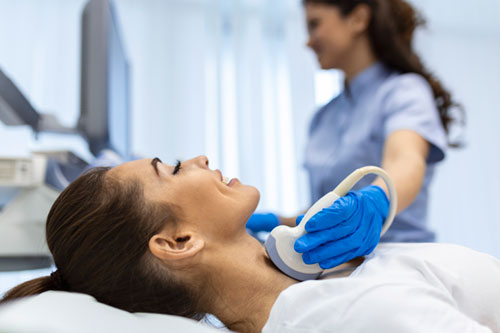
Thyroid sonography helps in the early detection of thyroid cancers, especially in populations with high risks like endemic goitre, radiation exposure, etc.
Carotid intima-media thickness (CIMT) measurement by ultrasound is proposed as a potential tool to aid cardiovascular risk stratification as it comprises a direct measure of atherosclerosis.
In extremely low birth weight and preterm babies < 32 weeks of gestational age, USG is used as a routine screening modality to detect the presence of germinal matrix and intraventricular neonatal haemorrhage.

Dr. Vijay
Senior Resident – Radiology,
Kauvery Hospital Chennai
